 Movie
Movie Controller
 Structure viewers
 About EMN search


-Search query
-Search result
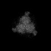
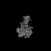
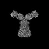
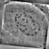
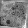
Showing 1 - 50 of 214 items for (author: schmid & mf)

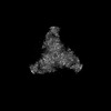
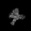
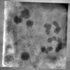
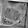
EMDB-49886: 
Cryo-ET map of the VZV capsid 3-fold axis.
Method: subtomogram averaging / : Oliver SL, Chen M

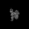
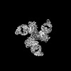
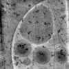
EMDB-49465:
Reconstruction of the intranuclear varicella-zoster virus capsid.
Method: subtomogram averaging / : Oliver SL

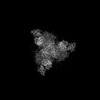
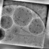
EMDB-49466:
Reconstruction of the varicella-zoster virus capsid vertex.
Method: subtomogram averaging / : Olver SL

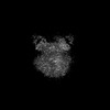
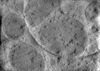
EMDB-49467:
Reconstruction of the intracellular varicella-zoster virus capsid with portal.
Method: subtomogram averaging / : Oliver SL

EMDB-49468:
VZV portal vertex cryo-ET reconstruction.
Method: subtomogram averaging / : Oliver SL

EMDB-49469:
VZV portal cryo-ET reconstruction.
Method: subtomogram averaging / : Oliver SL

EMDB-49470:
Reconstruction of intracellular varicella zoster virus CAI-capsid with portal.
Method: subtomogram averaging / : Oliver SL

EMDB-49471:
Reconstruction of the portal vertex from intracellular varicella-zoster virus CAI-capsids.
Method: subtomogram averaging / : Oliver SL

EMDB-49472:
Reconstruction of the varicella-zoster virus C-capsid with the portal vertex.
Method: subtomogram averaging / : Oliver SL

EMDB-49473:
VZV C-capsid portal vertex.
Method: subtomogram averaging / : Oliver SL

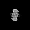
EMDB-47463:
Apo TRiC in closed conformation
Method: single particle / : Zhao Y, Chiu W

EMDB-47464:
TRiC encapsulating tubulin folding nucleus state.
Method: single particle / : Zhao Y, Chiu W

EMDB-47465:
Tubulin intermediate state I in human chaperonin TRiC
Method: single particle / : Zhao Y, Chiu W

EMDB-47466:
Tubulin intermediate state II in chaperonin TRiC
Method: single particle / : Zhao Y, Chiu W

EMDB-47467:
Tubulin intermediate III in chaperonin TRiC
Method: single particle / : Zhao Y, Chiu W

EMDB-47468:
Native Tubulin in human chaperonin TRIC
Method: single particle / : Zhao Y, Chiu W

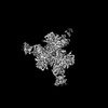
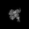
EMDB-49591:
Cryo-ET map of the VZV capsid vertex (5-fold axis).
Method: subtomogram averaging / : Oliver SL, Chen M

EMDB-45793:
Closed state MmCpn under ATP/AlFx condition by subtomogram averaging
Method: subtomogram averaging / : Yanyan Z

EMDB-45794:
MmCpn in closed state wit D9 symmetry
Method: subtomogram averaging / : Yanyan Z

EMDB-44925:
Structure of the LM189-Bound beta2AR-Gi Complex
Method: single particle / : Casiraghi M, Wang H, Kobilka BK
EMDB-41346:
CRYO-EM STRUCTURE OF HIV-1 BG505DS-SOSIP.664 ENV TRIMER BOUND TO DJ85-b.01 FAB
Method: single particle / : Pletnev S, Hoyt F, Fischer E, Kwong P
EMDB-41359:
CRYO-EM STRUCTURE OF HIV-1 BG505DS-SOSIP.664 ENV TRIMER BOUND TO DJ85-c.01 FAB
Method: single particle / : Pletnev S, Hoyt F, Fischer E, Kwong P
EMDB-41360:
CRYO-EM STRUCTURE OF HIV-1 BG505DS-SOSIP.664 ENV TRIMER BOUND TO DJ85-d.01 FAB
Method: single particle / : Pletnev S, Hoyt F, Fischer E, Kwong P
EMDB-41361:
CRYO-EM STRUCTURE OF HIV-1 BG505DS-SOSIP.664 ENV TRIMER BOUND TO DJ85-e.01 FAB
Method: single particle / : Pletnev S, Hoyt F, Fischer E, Kwong P
EMDB-41362:
CRYO-EM STRUCTURE OF HIV-1 BG505DS-SOSIP.664 ENV TRIMER BOUND TO HERH-c.01 FAB
Method: single particle / : Pletnev S, Hoyt F, Fischer E, Kwong P
EMDB-41426:
Cryo-EM structure of TRNM-b*01 Fab in complex with HIV-1 Env trimer BG505.DS SOSIP
Method: single particle / : Roark RS, Morano NC, Shapiro LS, Kwong PD

EMDB-41438:
Cryo-EM structure of HERH-b*01 Fab in complex with HIV-1 Env trimer BG505.DS SOSIP
Method: single particle / : Roark RS, Hoyt F, Hansen B, Fischer E, Shapiro LS, Kwong PD
EMDB-41440:
Cryo-EM structure of TRNM-f*01 Fab in complex with HIV-1 Env trimer ConC SOSIP
Method: single particle / : Roark RS, Morano NC, Shapiro LS, Kwong PD
EMDB-41459:
Cryo-EM structure of HIV-1 Env BG505 DS-SOSIP in complex with antibody GPZ6-b.01 targeting the fusion peptide
Method: single particle / : Zhou T, Morano NC, Roark RS, Kwong PD, Xu J
EMDB-41309:
CRYO-EM STRUCTURE OF HIV-1 BG505DS-SOSIP.664 ENV TRIMER BOUND TO HERH-c.01 FAB
Method: single particle / : Morano NC, Hoyt F, Hansen B, Fischer E, Shapiro L
EMDB-41310:
CRYO-EM STRUCTURE OF HIV-1 BG505DS-SOSIP.664 ENV TRIMER BOUND TO GPZ6-a.01 FAB
Method: single particle / : Morano NC, Becker JE, Shapiro L, Ho DD
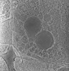
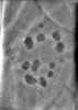
EMDB-28138:
3D reconstruction of the apical complex of Plasmodium falciparum (3D7) free merozoite
Method: electron tomography / : Segev-Zarko L, Sun SY, Kim CY
EMDB-28141:
3D reconstruction of the apical complex of Plasmodium falciparum (3D7) free merozoite
Method: electron tomography / : Segev-Zarko L, Sun SY, Kim CY

EMDB-28142:
3D Reconstruction of Plasmodium falciparum (3D7) free merozoite
Method: electron tomography / : Segev-Zarko L, Sun SY, Kim CY
EMDB-41840:
Gaussian Mixture Models based single particle refinement - GPCR (Substance P bound to active human neurokinin 1 receptor in complex with miniGs399 from EMPIAR-10786)
Method: single particle / : Chen M

EMDB-41841:
Gaussian mixture model based single particle refinement - SARS (SARS-CoV-2 Spike Proteins on intact virions from EMPIAR-10492)
Method: single particle / : Chen M
EMDB-41845:
Gaussian mixture model based single particle refinement - ABC transporter (inhibitor-bound ABCG2 from EMPIAR-10374)
Method: single particle / : Chen M

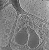
EMDB-28125:
Plasmodium falciparum merozoites apical 2-ring units
Method: subtomogram averaging / : Sun SY, Pintilie GD

EMDB-28126:
Toxoplasma apical rings
Method: subtomogram averaging / : Sun SY, Pintilie GD
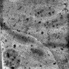
EMDB-29207:
CryoET tomogram of mitochondria in BACHD mouse model neuron
Method: electron tomography / : Wu GH, Galaz-Montoya JG, Gu Y, Mitchell PG, Wu C, Chiu W
EMDB-29208:
CryoET tomogram of BACHD mouse model neuron showing sheet aggregates
Method: electron tomography / : Wu GH, Galaz-Montoya JG, Gu Y, Mitchell PG, Wu C, Chiu W
EMDB-29210:
CryoET tomogram of purified mitochondria from HD patient iPSC-derived neuron (Q109)
Method: electron tomography / : Wu GH, Smith-Geater C, Galaz-Montoya JG, Mitchell PG, Thompson LM, Chiu W
EMDB-29211:
CryoET tomogram of HD patient iPSC-derived neuron (Q66) with PIAS1 hetKO treatment
Method: electron tomography / : Wu GH, Smith-Geater C, Galaz-Montoya JG, Mitchell PG, Thompson LM, Chiu W
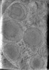
EMDB-28668:
CryoET tomogram of iPSC-derived control non-HD neuron (Q18)
Method: electron tomography / : Wu GH, Smith-Geater C, Galaz-Montoya JG, Mitchell PG, Thompson LM, Chiu W
EMDB-28944:
CryoET tomogram of iPSC-derived control non-HD neuron (Q20)
Method: electron tomography / : Wu GH, Smith-Geater C, Galaz-Montoya JG, Mitchell PG, Thompson LM, Chiu W
EMDB-28946:
CryoET tomogram of HD patient iPSC-derived neuron (Q53)
Method: electron tomography / : Wu GH, Smith-Geater C, Galaz-Montoya JG, Mitchell PG, Thompson LM, Chiu W
EMDB-29074:
CryoET tomogram of HD patient iPSC-derived neuron (Q66)
Method: electron tomography / : Wu GH, Smith-Geater C, Galaz-Montoya JG, Mitchell PG, Thompson LM, Chiu W
EMDB-29075:
CryoET tomogram of HD patient iPSC-derived neuron (Q77)
Method: electron tomography / : Wu GH, Smith-Geater C, Galaz-Montoya JG, Mitchell PG, Thompson LM, Chiu W
EMDB-29076:
CryoET tomogram of HD patient iPSC-derived neuron (Q109)
Method: electron tomography / : Wu GH, Smith-Geater C, Galaz-Montoya JG, Mitchell PG, Thompson LM, Chiu W
EMDB-29079:
CryoET tomogram of HD patient iPSC-derived neuron (Q66) showing sheet aggregate
Method: electron tomography / : Wu GH, Smith-Geater C, Galaz-Montoya JG, Mitchell PG, Thompson LM, Chiu W
Pages:
 wwPDB to switch to version 3 of the EMDB data model
