 Movie
Movie Controller
 Structure viewers
 About EMN search


-Search query
-Search result
Showing 1 - 50 of 116 items for (author: rogers & s)

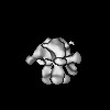
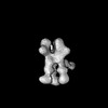
EMDB-49997: 
DHIK wk12 + Rhesus Macaque polyFab
Method: single particle / : Lin RN, Ward AB

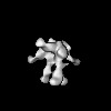
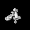
EMDB-70024:
Rhesus Macaque mAb CHM-27 complexed with SARS-CoV-2 spike protein
Method: single particle / : Lin RN, Ward AB

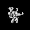
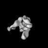
EMDB-70025:
Rhesus Macaque mAb CHM-16 complexed with SARS-CoV-2 spike protein
Method: single particle / : Lin RN, Ward AB

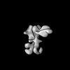
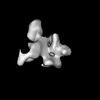
EMDB-70026:
Rhesus Macaque DHIK wk40 polyFab + SARS-CoV-2 Spike
Method: single particle / : Lin RN, Ward AB

EMDB-70027:
Rhesus Macaque DHJB wk12 polyFab + SARS-CoV-2 Spike
Method: single particle / : Lin RN, Ward AB

EMDB-70028:
Rhesus Macaque L603 wk53 polyFab + SARS-CoV-2 Spike
Method: single particle / : Lin RN, Ward AB

EMDB-70029:
Rhesus Macaque L603 wk40 polyFab + SARS-CoV-2 Spike
Method: single particle / : Lin RN, Ward AB

EMDB-70030:
Rhesus Macaque DHJB wk40 polyFab + SARS-CoV-2 Spike
Method: single particle / : Lin RN, Ward AB

EMDB-70031:
Rhesus Macaque L603 wk12 polyFab + SARS-CoV-2 Spike
Method: single particle / : Lin RN, Ward AB

EMDB-70032:
Rhesus Macaque K620 wk12 polyFab + SARS-CoV-2 Spike
Method: single particle / : Lin RN, Ward AB

EMDB-70033:
Rhesus Macaque K620 wk53 polyFab + SARS-CoV-2 Spike
Method: single particle / : Lin RN, Ward AB

EMDB-70034:
Rhesus Macaque K620 wk40 polyFab + SARS-CoV-2 Spike
Method: single particle / : Lin RN, Ward AB

EMDB-70624:
Cryo-EM structure of an octameric RAD51-XRCC3-RAD51C (RAD51-X3C) complex
Method: single particle / : Jia L, Ruben EA, Olsen SK, Wasmuth EV, Rawal Y, Kwon Y, Sung P

EMDB-70625:
Cryo-EM structure of a pentameric RAD51-XRCC3-RAD51C-RAD51D-XRCC2 (51-X3CDX2) complex.
Method: single particle / : Ruben EA, Jia L, Olsen SK, Wasmuth EV, Rawal Y, Kwon Y, Sung P

EMDB-70627:
Cryo-EM structure of a tetrameric XRCC3-RAD51C-RAD51D-XRCC2 complex
Method: single particle / : Ruben EA, Jia L, Olsen SK, Wasmuth EV, Rawal Y, Kwon Y, Sung P

EMDB-75014:
Cryo-EM Structure of a RAD51 filament bound by ssDNA and the XRCC3-RAD51C-RAD51D-XRCC2 paralog complex
Method: single particle / : Ruben EA, Jia L, Olsen SK, Wasmuth EV, Rawal Y, Kwon Y, Sung P

PDB-9omy:
Cryo-EM structure of an octameric RAD51-XRCC3-RAD51C (RAD51-X3C) complex
Method: single particle / : Jia L, Ruben EA, Olsen SK, Wasmuth EV, Rawal Y, Kwon Y, Sung P

PDB-9omz:
Cryo-EM structure of a pentameric RAD51-XRCC3-RAD51C-RAD51D-XRCC2 (51-X3CDX2) complex.
Method: single particle / : Ruben EA, Jia L, Olsen SK, Wasmuth EV, Rawal Y, Kwon Y, Sung P

PDB-9on2:
Cryo-EM structure of a tetrameric XRCC3-RAD51C-RAD51D-XRCC2 complex
Method: single particle / : Ruben EA, Jia L, Olsen SK, Wasmuth EV, Rawal Y, Kwon Y, Sung P

PDB-9zzr:
Cryo-EM Structure of a RAD51 filament bound by ssDNA and the XRCC3-RAD51C-RAD51D-XRCC2 paralog complex
Method: single particle / : Ruben EA, Jia L, Olsen SK, Wasmuth EV, Rawal Y, Kwon Y, Sung P

EMDB-52852:
structure of two human ELF2 transcription factors in complex with a nucleosome
Method: single particle / : Xiao T, Crowe-McAuliffe C, Dienemann C, Taipale J

EMDB-49952:
Dimerized subunit pair: modular DNA origami subunit with top = 14 poly-T, bottom = 3 poly-T overhangs
Method: single particle / : Videbaek TE, Wei WS, Rogers WB, Fraden S

EMDB-49953:
Dimerized subunit pair: modular DNA origami subunit with top = 3 poly-T, bottom = 3 poly-T overhangs
Method: single particle / : Videbaek TE, Wei WS, Rogers WB, Fraden S

EMDB-48565:
Modular DNA origami building block for self-assembly: dimer with 10 base pair arm
Method: single particle / : Videbaek TE, Hayakawa D, Saha R, Rogers WB, Fraden S

EMDB-48566:
Modular DNA origami building block for self-assembly: dimer with 6 base pair arm
Method: single particle / : Videbaek TE, Hayakawa D, Saha R, Rogers WB, Fraden S

EMDB-48567:
Modular DNA origami building block for self-assembly: dimer with 10 base pair arm, negative binding angle
Method: single particle / : Videbaek TE, Hayakawa D, Saha R, Rogers WB, Fraden S

EMDB-48569:
Modular DNA origami building block for self-assembly
Method: single particle / : Videbaek TE, Hayakawa D, Saha R, Rogers WB, Fraden S

EMDB-48633:
DNA-origami colloid for self-assembly of tubules: (10,0) monomer, bound pair from side 1 interaction
Method: single particle / : Videbaek TE, Rogers WB

EMDB-48634:
DNA-origami colloid for self-assembly of tubules: (10,0) monomer, bound pair from side 2 interaction
Method: single particle / : Videbaek TE, Rogers WB

EMDB-48635:
DNA-origami colloid for self-assembly of tubules: (10,0) monomer, bound pair from side 3 interaction
Method: single particle / : Videbaek TE, Rogers WB

EMDB-48636:
DNA-origami colloid for self-assembly of tubules: (10,0) monomer, bound hexamer
Method: single particle / : Videbaek TE, Rogers WB

EMDB-48637:
DNA-origami colloid for self-assembly of tubules: (10,0) monomer, twist corrected
Method: single particle / : Videbaek TE, Rogers WB
EMDB-41869:
BG505.664 SOSIP in complex with polyclonal antibodies from NHP 8131 (gp120 glycan hole, gp41 glycan hole/fusion peptide and trimer base epitopes)
Method: single particle / : Zhang S, Ward AB
EMDB-41870:
BG505.664 Env SOSIP in complex with polyclonal antibodies from NHP 8131 (gp120-gp120 interface epitope)
Method: single particle / : Zhang S, Ward AB

EMDB-41871:
BG505.664 Env SOSIP in complex with polyclonal antibodies from NHP 8147 (C3/V5, V1/V2/V3 apex, gp41 glycan hole/fusion peptide and trimer base epitopes)
Method: single particle / : Zhang S, Ward AB
EMDB-41872:
BG505.664 Env SOSIP in complex with polyclonal antibodies from NHP 8147 (gp120 glycan hole epitope)
Method: single particle / : Zhang S, Ward AB
EMDB-41972:
GT1.1 SOSIP in complex with wk39 polyclonal antibodies from NHP A12N030 (CD4bs, C3V5 and base epitopes)
Method: single particle / : van Schooten J, Ozorowski G, Ward AB
EMDB-41973:
GT1.1 SOSIP in complex with wk39 polyclonal antibodies from NHP A12N030 (gp41GH/FP and base epitopes)
Method: single particle / : van Schooten J, Ozorowski G, Ward AB
EMDB-41974:
GT1.1 SOSIP in complex with wk39 polyclonal antibodies from NHP DC8G (gp41GH/FP and gp120GH epitopes)
Method: single particle / : van Schooten J, Ozorowski G, Ward AB
EMDB-41975:
GT1.1 SOSIP in complex with wk39 polyclonal antibodies from NHP DC8G (gp120GH and base epitopes)
Method: single particle / : van Schooten J, Ozorowski G, Ward AB
EMDB-41976:
GT1.1 SOSIP in complex with wk39 polyclonal antibodies from NHP DC8G (V1V2V3 and gp120GH epitopes)
Method: single particle / : van Schooten J, Ozorowski G, Ward AB
EMDB-41977:
GT1.1 SOSIP-CC2 in complex with wk80 polyclonal antibodies from NHP 8229 (V1V2V3, C3V5, CD4bs, gp41GH/FP and base epitopes)
Method: single particle / : Sewell LM, Ozorowski G, Ward AB
EMDB-41978:
GT1.1 SOSIP-CC2 in complex with wk80 polyclonal antibodies from NHP 8239 (gp120GH, gp41GH/FP, CD4bs, and base epitopes)
Method: single particle / : Sewell LM, Ozorowski G, Ward AB

EMDB-40796:
BG505 GT1.1 SOSIP in complex with NHP Fabs 12C11 and RM20A3
Method: single particle / : Zhang S, Torres JL, Ozorowski G, Ward AB

PDB-8sw3:
BG505 GT1.1 SOSIP in complex with NHP Fabs 12C11 and RM20A3
Method: single particle / : Zhang S, Torres JL, Ozorowski G, Ward AB

EMDB-40812:
Structure of SARS-CoV-2 (HP-GSAS-Mut7) spike in complex with TXG-0078 Fab -Conformation 1
Method: single particle / : Bangaru S, Ward AB

EMDB-40813:
Structure of SARS-CoV-2 (HP-GSAS-Mut7) spike in complex with TXG-0078 Fab -Conformation 2
Method: single particle / : Bangaru S, Ward AB
EMDB-40797:
BG505 GT1.1 SOSIP in complex with NHP Fabs 21N13, 21M20 and RM20A3
Method: single particle / : Ozorowski G, Torres JL, Zhang S, Ward AB

PDB-8sw4:
BG505 GT1.1 SOSIP in complex with NHP Fabs 21N13, 21M20 and RM20A3
Method: single particle / : Ozorowski G, Torres JL, Zhang S, Ward AB

EMDB-40814:
Local refinement of SARS-CoV-2 (HP-GSAS-Mut7) spike NTD in complex with TXG-0078 Fab
Method: single particle / : Bangaru B, Ward A
Pages:
 wwPDB to switch to version 3 of the EMDB data model
