 Movie
Movie Controller

[English] 日本語
 Yorodumi
- PDB-8oh4: Subtomogram averaging structure of cofilactin filament inside mic... -
+ Open data

- Basic information
| Entry | Database: PDB / ID: 8oh4 | |||||||||
|---|---|---|---|---|---|---|---|---|---|---|
| Title | Subtomogram averaging structure of cofilactin filament inside microtubule lumen of Drosophila S2 cell protrusion. | |||||||||
 Components Components |
| |||||||||
 Keywords Keywords | CONTRACTILE PROTEIN / Cytoskeleton / Filament / Actin / Cofilactin | |||||||||
| Function / homology |  Function and homology information Function and homology informationestablishment of imaginal disc-derived wing hair orientation / establishment of ommatidial planar polarity / imaginal disc-derived leg segmentation / Gap junction degradation / Formation of annular gap junctions / EPHB-mediated forward signaling / EPH-ephrin mediated repulsion of cells / Recycling pathway of L1 / Cell-extracellular matrix interactions / RHOBTB2 GTPase cycle ...establishment of imaginal disc-derived wing hair orientation / establishment of ommatidial planar polarity / imaginal disc-derived leg segmentation / Gap junction degradation / Formation of annular gap junctions / EPHB-mediated forward signaling / EPH-ephrin mediated repulsion of cells / Recycling pathway of L1 / Cell-extracellular matrix interactions / RHOBTB2 GTPase cycle / RHOF GTPase cycle / Regulation of CDH1 Function / Formation of the canonical BAF (cBAF) complex / Formation of the polybromo-BAF (pBAF) complex / Formation of the embryonic stem cell BAF (esBAF) complex / Formation of the non-canonical BAF (ncBAF) complex / Formation of neuronal progenitor and neuronal BAF (npBAF and nBAF) / VEGFA-VEGFR2 Pathway / ovarian fusome organization / Interaction between L1 and Ankyrins / Platelet degranulation / RHO GTPases Activate WASPs and WAVEs / Regulation of actin dynamics for phagocytic cup formation / Formation of the dystrophin-glycoprotein complex (DGC) / compound eye morphogenesis / DNA Damage Recognition in GG-NER / rhabdomere development / meiotic cytokinesis / UCH proteinases / mushroom body development / actomyosin contractile ring assembly / Clathrin-mediated endocytosis / border follicle cell migration / compound eye development / sperm individualization / establishment of planar polarity / centrosome separation / actin filament fragmentation / epithelial structure maintenance / maintenance of protein location in cell / brahma complex / tube formation / actin filament severing / regulation of lamellipodium assembly / actin filament depolymerization / Ino80 complex / lamellipodium assembly / female gonad development / mitotic cytokinesis / axonogenesis / actin filament polymerization / actin filament organization / positive regulation of protein secretion / Hydrolases; Acting on acid anhydrides; Acting on acid anhydrides to facilitate cellular and subcellular movement / structural constituent of cytoskeleton / nuclear matrix / actin filament binding / actin cytoskeleton / actin binding / chromatin remodeling / ATP hydrolysis activity / nucleoplasm / ATP binding / nucleus / cytosol / cytoplasm Similarity search - Function | |||||||||
| Biological species |  | |||||||||
| Method | ELECTRON MICROSCOPY / subtomogram averaging / cryo EM / Resolution: 16.5 Å | |||||||||
 Authors Authors | Ventura Santos, C. / Carter, A.P. | |||||||||
| Funding support |  United Kingdom, 2items United Kingdom, 2items
| |||||||||
 Citation Citation | Journal: bioRxiv / Year: 2023 Title: CryoET shows cofilactin filaments inside the microtubule lumen. Authors: Camilla Ventura Santos / Stephen L Rogers / Andrew P Carter / Abstract: Cytoplasmic microtubules are tubular polymers that can harbor small proteins or filaments inside their lumen. The identity of these objects and what causes their accumulation has not been ...Cytoplasmic microtubules are tubular polymers that can harbor small proteins or filaments inside their lumen. The identity of these objects and what causes their accumulation has not been conclusively established. Here, we used cryogenic electron tomography (cryoET) of S2 cell protrusions and found filaments inside the microtubule lumen, which resemble those reported recently in human HAP1 cells. The frequency of these filaments increased upon inhibition of the sarco/endoplasmic reticulum Ca ATPase (SERCA) with the small-molecule drug thapsigargin. Subtomogram averaging showed that the luminal filaments adopt a helical structure reminiscent of cofilin-bound actin (cofilactin). Consistent with this, cofilin was activated in cells under the same conditions that increased luminal filament occurrence. Furthermore, RNAi knock-down of cofilin reduced the frequency of luminal filaments with cofilactin morphology. These results suggest that cofilin activation stimulates its accumulation on actin filaments inside the microtubule lumen. | |||||||||
| History |
|
- Structure visualization
| Structure viewer | Molecule: MolmilJmol/JSmol |
|---|
- Downloads & links
-Download
| PDBx/mmCIF format | 8oh4.cif.gz | 458.6 KB | Display | PDBx/mmCIF format |
|---|---|---|---|---|
| PDB format | pdb8oh4.ent.gz | 302.8 KB | Display | PDB format |
| PDBx/mmJSON format | 8oh4.json.gz | Tree view | PDBx/mmJSON format | |
| Others |  Other downloads Other downloads |
-Validation report
| Arichive directory | https://data.pdbj.org/pub/pdb/validation_reports/oh/8oh4ftp://data.pdbj.org/pub/pdb/validation_reports/oh/8oh4 | HTTPS FTP |
|---|
-Related structure data
| Related structure data | 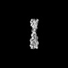 16877MC M: map data used to model this data C: citing same article ( |
|---|---|
| Similar structure data |
-Links
 PDBj
PDBj


- Assembly
| Deposited unit | 
|
|---|---|
| 1 |
|
-Components
| #1: Protein | Mass: 41160.980 Da / Num. of mol.: 8 Mutation: 6 N-terminal residues (MCDEEV) were removed. Residues 42-50 (QGVMVGMGC) were removed Source method: isolated from a natural source / Source: (natural) References: UniProt: P10987, Hydrolases; Acting on acid anhydrides; Acting on acid anhydrides to facilitate cellular and subcellular movement #2: Protein | Mass: 17180.529 Da / Num. of mol.: 6 / Source method: isolated from a natural source / Source: (natural) |
|---|
-Experimental details
-Experiment
| Experiment | Method: ELECTRON MICROSCOPY |
|---|---|
| EM experiment | Aggregation state: FILAMENT / 3D reconstruction method: subtomogram averaging |
- Sample preparation
| Component | Name: Cofilactin filament inside the microtubule lumen of induced Drosophila S2 cell protrusion Type: CELL / Entity ID: all / Source: NATURAL |
|---|---|
| Source (natural) | Organism: |
| Buffer solution | pH: 7 |
| Specimen | Embedding applied: NO / Shadowing applied: NO / Staining applied: NO / Vitrification applied: YES |
| Specimen support | Details: Quantifoil R3.5/1 Au200 grids were glow discharged for 30s at 20 - 30 mA and subsequently coated with 0.25 ug/mL Concanavalin Aater for 1 - 16 h at 37 degrees Celcius. Grid material: GOLD / Grid mesh size: 200 divisions/in. / Grid type: Quantifoil R3.5/1 |
| Vitrification | Instrument: FEI VITROBOT MARK III / Cryogen name: ETHANE / Humidity: 95 % / Chamber temperature: 198.15 K |
- Electron microscopy imaging
| Experimental equipment |  Model: Titan Krios / Image courtesy: FEI Company |
|---|---|
| Microscopy | Model: FEI TITAN KRIOS |
| Electron gun | Electron source:  FIELD EMISSION GUN / Accelerating voltage: 300 kV / Illumination mode: FLOOD BEAM FIELD EMISSION GUN / Accelerating voltage: 300 kV / Illumination mode: FLOOD BEAM |
| Electron lens | Mode: BRIGHT FIELD / Nominal defocus max: 6000 nm / Nominal defocus min: 2500 nm |
| Image recording | Electron dose: 3 e/Å2 / Avg electron dose per subtomogram: 120 e/Å2 / Detector mode: COUNTING / Film or detector model: GATAN K2 SUMMIT (4k x 4k) Details: Data was collected on Gatan K2 summit (2.952 A/pixel) and Gatan K3 summit (2.659 A/pixel) with 3 degree increments (3 e/A2 dose per tilt). The total dose was between 118 and 122 e/A2. |
| EM imaging optics | Energyfilter name: GIF Bioquantum / Energyfilter slit width: 20 eV |
- Processing
| EM software |
| ||||||||||||||||
|---|---|---|---|---|---|---|---|---|---|---|---|---|---|---|---|---|---|
| CTF correction | Details: CTF estimation was performed in WARP. CTF correction was performed in Relion 3.1. Type: PHASE FLIPPING AND AMPLITUDE CORRECTION | ||||||||||||||||
| Symmetry | Point symmetry: C1 (asymmetric) | ||||||||||||||||
| 3D reconstruction | Resolution: 16.5 Å / Resolution method: FSC 0.143 CUT-OFF / Num. of particles: 3801 Details: helical symmetry (-162 twist, 29A rise) was applied during 3D classification but not during refinements. Symmetry type: POINT | ||||||||||||||||
| EM volume selection | Num. of tomograms: 58 / Num. of volumes extracted: 7549 | ||||||||||||||||
| Atomic model building | Details: Drosophila cofilin and actin structures were predicted as a complex with Alphafold 2-Multimer. Actin from the predicted cofilin-actin complex was iteratively aligned with 8 actin subunits in ...Details: Drosophila cofilin and actin structures were predicted as a complex with Alphafold 2-Multimer. Actin from the predicted cofilin-actin complex was iteratively aligned with 8 actin subunits in the chicken cofilactin PDB model 5yU8. Two cofilin moieties at the pointed end were removed. Side chains were truncated. The model was not refined. |
