 Movie
Movie Controller

[English] 日本語
 Yorodumi
- EMDB-24764: in situ cryo-electron tomogram of wild-type yeast lipid droplet a... -
+ Open data

- Basic information
| Entry |  | |||||||||||||||
|---|---|---|---|---|---|---|---|---|---|---|---|---|---|---|---|---|
| Title | in situ cryo-electron tomogram of wild-type yeast lipid droplet after 4-hour exposure to acute glucose restriction (AGR, 0.001% glucose media) | |||||||||||||||
 Map data Map data | ||||||||||||||||
 Sample Sample |
| |||||||||||||||
| Biological species |  | |||||||||||||||
| Method | electron tomography / cryo EM | |||||||||||||||
 Authors Authors | Rogers S / Gui L / Kovalenko A / Reetz E / Nicastro D / Henne WM | |||||||||||||||
| Funding support |  United States, 4 items United States, 4 items
| |||||||||||||||
 Citation Citation | Journal: To Be Published Title: Liquid-crystalline lipid phase transitions in lipid droplets selectively remodel the LD proteome Authors: Rogers S / Gui L / Kovalenko A / Reetz E / Nicastro D / Henne WM | |||||||||||||||
| History |
|
- Structure visualization
| Supplemental images |
|---|

- Downloads & links
-EMDB archive
| Map data | emd_24764.map.gz | 100.4 MB |  EMDB map data format EMDB map data format | |
|---|---|---|---|---|
| Header (meta data) | emd-24764-v30.xmlemd-24764.xml | 8.2 KB 8.2 KB | Display Display | EMDB header |
| Images | 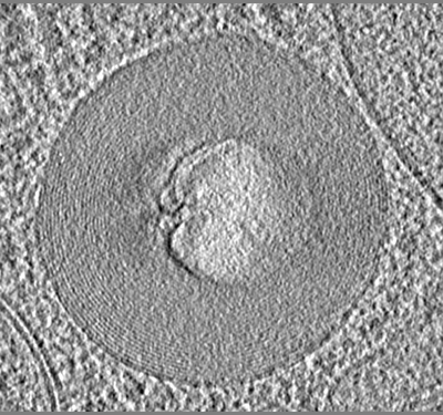 emd_24764.png emd_24764.png | 182.9 KB | ||
| Archive directory |  http://ftp.pdbj.org/pub/emdb/structures/EMD-24764ftp://ftp.pdbj.org/pub/emdb/structures/EMD-24764 http://ftp.pdbj.org/pub/emdb/structures/EMD-24764ftp://ftp.pdbj.org/pub/emdb/structures/EMD-24764 | HTTPS FTP |
-Related structure data


-Links
| EMDB pages | EMDB (EBI/PDBe) / EMDataResource |
|---|
-Map
| File | Download / File: emd_24764.map.gz / Format: CCP4 / Size: 183.4 MB / Type: IMAGE STORED AS SIGNED BYTE | ||||||||||||||||||||
|---|---|---|---|---|---|---|---|---|---|---|---|---|---|---|---|---|---|---|---|---|---|
| Voxel size | X=Y=Z: 5.684 Å | ||||||||||||||||||||
| Density |
| ||||||||||||||||||||
| Symmetry | Space group: 1 | ||||||||||||||||||||
| Details | EMDB XML:
|
-Supplemental data
- Sample components
-Entire : lipid droplet of wild-type yeast after 4-hour exposure to acute g...
| Entire | Name: lipid droplet of wild-type yeast after 4-hour exposure to acute glucose restriction (AGR, 0.001% glucose media) |
|---|---|
| Components |
|
-Supramolecule #1: lipid droplet of wild-type yeast after 4-hour exposure to acute g...
| Supramolecule | Name: lipid droplet of wild-type yeast after 4-hour exposure to acute glucose restriction (AGR, 0.001% glucose media) type: cell / ID: 1 / Parent: 0 |
|---|---|
| Source (natural) | Organism: |
-Experimental details
-Structure determination
| Method | cryo EM |
|---|---|
 Processing Processing | electron tomography |
| Aggregation state | cell |
-Sample preparation
| Buffer | pH: 7 |
|---|---|
| Vitrification | Cryogen name: ETHANE |
| Sectioning | Focused ion beam - Instrument: OTHER / Focused ion beam - Ion: OTHER / Focused ion beam - Voltage: 30 kV / Focused ion beam - Current: 0.01 nA / Focused ion beam - Duration: 600 sec. / Focused ion beam - Temperature: 88 K / Focused ion beam - Initial thickness: 1000 nm / Focused ion beam - Final thickness: 150 nm Focused ion beam - Details: The value given for _emd_sectioning_focused_ion_beam.instrument is FEI Aquilos. This is not in a list of allowed values {'DB235', 'OTHER'} so OTHER is written into the XML file. |
- Electron microscopy
| Microscope | FEI TITAN KRIOS |
|---|---|
| Image recording | Film or detector model: GATAN K3 (6k x 4k) / Average electron dose: 1.5 e/Å2 |
| Electron beam | Acceleration voltage: 300 kV / Electron source:  FIELD EMISSION GUN FIELD EMISSION GUN |
| Electron optics | Illumination mode: FLOOD BEAM / Imaging mode: BRIGHT FIELD |
| Experimental equipment |  Model: Titan Krios / Image courtesy: FEI Company |
-Image processing
| Final reconstruction | Number images used: 57 |
|---|
