 Movie
Movie Controller
 Structure viewers
 About EMN search


-Search query
-Search result
Showing 1 - 50 of 62 items for (author: pinto & jr)

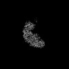
EMDB-48468: 
Structure of native murine cardiac thin filament at pCa=5.8 in Ca2+-free rotated state (upper strand)
Method: single particle / : Risi CM, Galkin VE

EMDB-48469:
Structure of native murine cardiac thin filament at pCa=5.8 in Ca2+-free tilted state (upper strand)
Method: single particle / : Risi CM, Galkin VE

EMDB-48470:
Structure of native murine cardiac thin filament at pCa=5.8 in Ca2+-bound partially activated state (upper strand)
Method: single particle / : Risi CM, Galkin VE

EMDB-48471:
Structure of native murine cardiac thin filament at pCa=5.8 in Ca2+-bound fully activated state (upper strand)
Method: single particle / : Risi CM, Galkin VE

EMDB-48476:
Structure of native murine cardiac thin filament variant I79N in troponin T at pCa=5.8 in Ca2+-free state (lower strand)
Method: single particle / : Risi CM, Galkin VE

EMDB-47449:
The structure of the junction region of the wild-type murine native cardiac thin filament in Ca2+-free state
Method: single particle / : Galkin VE, Risi CM

EMDB-48467:
Structure of native murine cardiac thin filament at pCa=5.8 in Ca2+-free rotated state (lower strand)
Method: single particle / : Galkin VE, Risi CM

EMDB-48477:
Structure of native murine cardiac thin filament variant I79N in troponin T at pCa=5.8 in Ca2+-bound fully activated state (lower strand)
Method: single particle / : Risi CM, Galkin VE

EMDB-48482:
Structure of native murine cardiac thin filament variant I79N in troponin T at pCa=5.8 in Ca2+-bound partially activated state (upper strand)
Method: single particle / : Risi CM, Galkin VE

EMDB-48483:
Structure of native murine cardiac thin filament variant I79N in troponin T at pCa=5.8 in Ca2+-bound fully activated state (upper strand)
Method: single particle / : Risi CM, Galkin VE

EMDB-48484:
Structure of native murine cardiac thin filament variant I79N in troponin T at pCa=5.8 in Ca2+-bound partially activated state (lower strand)
Method: single particle / : Risi CM, Galkin VE

EMDB-48447:
Structure of native murine cardiac thin filament at pCa=5.8 in Ca2+-free state (lower strand)
Method: single particle / : Risi CM, Galkin VE

EMDB-48448:
Structure of native murine cardiac thin filament at pCa=5.8 in Ca2+-bound partially activated state (lower strand)
Method: single particle / : Risi CM, Galkin VE

EMDB-48449:
Structure of native murine cardiac thin filament at pCa=5.8 in Ca2+-free tilted state (lower strand)
Method: single particle / : Risi CM, Galkin VE

EMDB-48450:
Structure of native murine cardiac thin filament at pCa=5.8 in Ca2+-bound fully activated state (lower strand)
Method: single particle / : Risi CM, Galkin VE

EMDB-48451:
Structure of native murine cardiac thin filament at pCa=5.8 in Ca2+-free state (upper strand)
Method: single particle / : Risi CM, Galkin VE

EMDB-48452:
Structure of native murine cardiac thin filament variant I79N in troponin T at pCa=5.8 in Ca2+-free rotated state (lower strand)
Method: single particle / : Risi CM, Galkin VE

EMDB-48453:
Structure of native murine cardiac thin filament variant I79N in troponin T at pCa=5.8 in Ca2+-free tilted state (lower strand)
Method: single particle / : Risi CM, Galkin VE

EMDB-48454:
Structure of native murine cardiac thin filament variant I79N in troponin T at pCa=5.8 in Ca2+-free state (upper strand)
Method: single particle / : Risi CM, Galkin VE

EMDB-48455:
Structure of native murine cardiac thin filament variant I79N in troponin T at pCa=5.8 in Ca2+-free rotated state (upper strand)
Method: single particle / : Risi CM, Galkin VE

EMDB-48456:
Structure of native murine cardiac thin filament variant I79N in troponin T at pCa=5.8 in Ca2+-free tilted state (upper strand)
Method: single particle / : Risi CM, Galkin VE
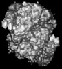
EMDB-43813:
VIR-7229 Fab fragment bound the SARS-CoV-2 BA.2.86 spike trimer (local refinement of the BA 2.86 RBD/VIR-7229 VHVL)
Method: single particle / : Park YJ, Tortorici MA, Seattle Structural Genomics Center for Infectious Disease (SSGCID), Veesler D
EMDB-43842:
VIR-7229 Fab fragment bound the BA.2.86 spike trimer (global refinement)
Method: single particle / : Tortorici MA, Park YJ, Veelser D, Seattle Structural Genomics Center for Infectious Disease (SSGCID)
EMDB-42681:
The structure of the native cardiac thin filament troponin core in Ca2+-free state from the upper strand
Method: single particle / : Galkin VE, Risi CM
EMDB-42682:
The structure of the native cardiac thin filament troponin core in Ca2+-free tilted state from the upper strand
Method: single particle / : Galkin VE, Risi CM

EMDB-42683:
The structure of the native cardiac thin filament troponin core in Ca2+-free rotated state from the upper strand
Method: single particle / : Galkin VE, Risi CM
EMDB-42800:
The structure of the native cardiac thin filament troponin core in Ca2+-free state from the lower strand
Method: single particle / : Galkin VE, Risi CM
EMDB-42833:
The structure of the native cardiac thin filament troponin core in Ca2+-free rotated state from the lower strand
Method: single particle / : Galkin VE, Risi CM
EMDB-42835:
The structure of the native cardiac thin filament troponin core in Ca2+-free tilted state from the lower strand
Method: single particle / : Galkin VE, Risi CM
EMDB-42846:
The structure of the native cardiac thin filament troponin core in Ca2+-bound fully activated state from the upper strand
Method: single particle / : Galkin VE, Risi CM

EMDB-42847:
The structure of the native cardiac thin filament troponin core in Ca2+-bound partially activated state from the upper strand
Method: single particle / : Galkin VE, Risi CM
EMDB-42849:
The structure of the native cardiac thin filament troponin core in Ca2+-bound fully activated state 1 from the lower strand
Method: single particle / : Galkin VE, Risi CM
EMDB-42856:
The structure of the native cardiac thin filament troponin core in Ca2+-bound fully activated state 2 from the lower strand
Method: single particle / : Galkin VE, Risi CM
EMDB-42858:
The structure of the native cardiac thin filament troponin core in Ca2+-bound partially activated state from the lower strand
Method: single particle / : Galkin VE, Risi CM

EMDB-42874:
The structure of the native cardiac thin filament troponin core in Ca2+-free state from the upper strand activated by the C1-domain of cardiac myosin binding protein C
Method: single particle / : Galkin VE, Risi CM

EMDB-40468:
In situ human cardiac thick filament in the relaxed state
Method: subtomogram averaging / : Chen L, Liu J, Rastegarpouyani H, Janssen PML, Pinto JR, Taylor KA

EMDB-40471:
Human cardiac interacting heads motif (IHM-C) in complete form
Method: subtomogram averaging / : Chen L, Liu J, Rastegarpouyani H, Janssen PML, Pinto JR, Taylor KA

EMDB-40475:
Human cardiac interacting heads motif (IHM-C) in semi form
Method: subtomogram averaging / : Chen L, Liu J, Rastegarpouyani H, Janssen PML, Pinto JR, Taylor KA

EMDB-40476:
Human cardiac interacting heads motif (IHM-S)
Method: subtomogram averaging / : Chen L, Liu J, Rastegarpouyani H, Janssen PML, Pinto JR, Taylor KA

EMDB-40478:
Human cardiac interacting heads motif (IHM-D) in semi form
Method: subtomogram averaging / : Chen L, Liu J, Rastegarpouyani H, Janssen PML, Pinto JR, Taylor KA
EMDB-29530:
SARS-CoV-2 XBB.1 spike RBD bound to the human ACE2 ectodomain and the S309 neutralizing antibody Fab fragment
Method: single particle / : Park YJ, Seattle Structural Genomics Center for Infectious Disease (SSGCID), Veesler D
EMDB-29531:
SARS-CoV-2 BQ.1.1 spike RBD bound to the human ACE2 ectodomain and the S309 neutralizing antibody Fab fragment
Method: single particle / : Park YJ, Seattle Structural Genomics Center for Infectious Disease (SSGCID), Veesler D

EMDB-40240:
SARS-CoV-2 BN.1 spike RBD bound to the human ACE2 ectodomain and the S309 neutralizing antibody Fab fragment
Method: single particle / : Park YJ, Seattle Structural Genomics Center for Infectious Disease (SSGCID), Veesler D
EMDB-27331:
The structure of the native cardiac thin filament junction region
Method: single particle / : Galkin VE, Risi CM
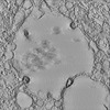
EMDB-14359:
Tomogram showing SARS-CoV-2 virions that have a budding like profile within a viral containing compartment
Method: electron tomography / : Pinto AL, Rai RK, Brown JC, Griffin P, Edgar JR, Shah A, Singanayagam A, Hogg C, Barclay WS, Futter CE, Burgoyne T

EMDB-14361:
Tomogram showing SARS-CoV-2 virions that have a budding like profile within a viral containing compartment
Method: electron tomography / : Pinto AL, Rai RK, Brown JC, Griffin P, Edgar JR, Shah A, Singanayagam A, Hogg C, Barclay WS, Futter CE, Burgoyne T
EMDB-14363:
Tomogram showing SARS-CoV-2 virions that have a budding like profile within a viral containing compartment
Method: electron tomography / : Pinto AL, Rai RK, Brown JC, Griffin P, Edgar JR, Shah A, Singanayagam A, Hogg C, Barclay WS, Futter CE, Burgoyne T
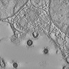
EMDB-14364:
Tomogram showing SARS-CoV-2 S glycoprotein on the membrane of a viral containing compartment
Method: electron tomography / : Pinto AL, Rai RK, Brown JC, Griffin P, Edgar JR, Shah A, Singanayagam A, Hogg C, Barclay WS, Futter CE, Burgoyne T
EMDB-14365:
Tomogram showing SARS-CoV-2 S glycoprotein on the membrane of a viral containing compartment
Method: electron tomography / : Pinto AL, Rai RK, Brown JC, Griffin P, Edgar JR, Shah A, Singanayagam A, Hogg C, Barclay WS, Futter CE, Burgoyne T
EMDB-14366:
Tomogram of a SARS-CoV-2 virion fused to the plasma membrane of a ciliated airway cell
Method: electron tomography / : Pinto AL, Rai RK, Brown JC, Griffin P, Edgar JR, Shah A, Singanayagam A, Hogg C, Barclay WS, Futter CE, Burgoyne T
Pages:
 wwPDB to switch to version 3 of the EMDB data model
