 Movie
Movie Controller
 Structure viewers
 About Yorodumi Papers


+Search query
-Structure paper
| Title | Ultrastructural insight into SARS-CoV-2 entry and budding in human airway epithelium. |
|---|---|
| Journal, issue, pages | Nat Commun, Vol. 13, Issue 1, Page 1609, Year 2022 |
| Publish date | Mar 25, 2022 |
 Authors Authors | Andreia L Pinto / Ranjit K Rai / Jonathan C Brown / Paul Griffin / James R Edgar / Anand Shah / Aran Singanayagam / Claire Hogg / Wendy S Barclay / Clare E Futter / Thomas Burgoyne /  |
| PubMed Abstract | Ultrastructural studies of SARS-CoV-2 infected cells are crucial to better understand the mechanisms of viral entry and budding within host cells. Here, we examined human airway epithelium infected ...Ultrastructural studies of SARS-CoV-2 infected cells are crucial to better understand the mechanisms of viral entry and budding within host cells. Here, we examined human airway epithelium infected with three different isolates of SARS-CoV-2 including the B.1.1.7 variant by transmission electron microscopy and tomography. For all isolates, the virus infected ciliated but not goblet epithelial cells. Key SARS-CoV-2 entry molecules, ACE2 and TMPRSS2, were found to be localised to the plasma membrane including microvilli but excluded from cilia. Consistently, extracellular virions were seen associated with microvilli and the apical plasma membrane but rarely with ciliary membranes. Profiles indicative of viral fusion where tomography showed that the viral membrane was continuous with the apical plasma membrane and the nucleocapsids diluted, compared with unfused virus, demonstrate that the plasma membrane is one site of entry where direct fusion releasing the nucleoprotein-encapsidated genome occurs. Intact intracellular virions were found within ciliated cells in compartments with a single membrane bearing S glycoprotein. Tomography showed concentration of nucleocapsids round the periphery of profiles strongly suggestive of viral budding into these compartments and this may explain how virions gain their S glycoprotein containing envelope. |
 External links External links | Nat Commun / PubMed:35338134 / PubMed Central |
| Methods | EM (tomography) |
| Structure data | 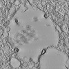 EMDB-14359: Tomogram showing SARS-CoV-2 virions that have a budding like profile within a viral containing compartment  EMDB-14361: Tomogram showing SARS-CoV-2 virions that have a budding like profile within a viral containing compartment 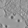 EMDB-14363: Tomogram showing SARS-CoV-2 virions that have a budding like profile within a viral containing compartment 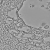 EMDB-14364: Tomogram showing SARS-CoV-2 S glycoprotein on the membrane of a viral containing compartment 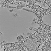 EMDB-14365: Tomogram showing SARS-CoV-2 S glycoprotein on the membrane of a viral containing compartment 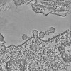 EMDB-14366: Tomogram of a SARS-CoV-2 virion fused to the plasma membrane of a ciliated airway cell 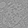 EMDB-14367: Tomogram showing SARS-CoV-2 virions within a viral containing compartment of an infected airway cell |
| Source |
|

 Severe acute respiratory syndrome coronavirus 2