 ムービー
ムービー コントローラー
 構造ビューア
 EMN検索について


-検索条件
-検索結果
検索 (著者・登録者: miki & m)の結果152件中、1から50件目までを表示しています
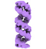
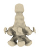
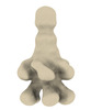
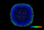
EMDB-19599: 
Structural characterization of Thogoto Virus nucleoprotein provides insights into RNA encapsidation and assembly
PDB-8ryt:
Structural characterization of Thogoto Virus nucleoprotein provides insights into RNA encapsidation and assembly

EMDB-38453:
Structure of the SARS-CoV-2 EG.5.1 spike glycoprotein in complex with ACE2 (1-up state)

EMDB-38454:
Structure of the SARS-CoV-2 EG.5.1 spike RBD in complex with ACE2

PDB-8xlm:
Structure of the SARS-CoV-2 EG.5.1 spike glycoprotein in complex with ACE2 (1-up state)

PDB-8xln:
Structure of the SARS-CoV-2 EG.5.1 spike RBD in complex with ACE2

EMDB-37648:
SARS-CoV-2 EG.5.1 spike glycoprotein (1-up state)

EMDB-37650:
SARS-CoV-2 EG.5.1 spike glycoprotein (closed-2 state)

EMDB-37651:
SARS-CoV-2 EG.5.1 spike glycoprotein (closed-1 state)

PDB-8wmd:
Structure of the SARS-CoV-2 EG.5.1 spike glycoprotein (closed-2 state)

PDB-8wmf:
Structure of the SARS-CoV-2 EG.5.1 spike glycoprotein (closed-1 state)
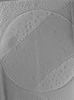
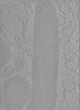
EMDB-18953:
Cryo-electron tomogram of Yersinia entomophaga chi2-sfGFP cells
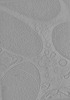
EMDB-18954:
Cryo-electron tomogram of Yersinia entomophaga chi2-sfGFP cells
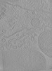
EMDB-18955:
Cryo-electron tomogram of Yersinia entomophaga chi2-sfGFP cells
EMDB-18957:
Cryo-electron tomogram of Yersinia entomophaga MH96 cells
EMDB-18958:
Cryo-electron tomogram of Yersinia entomophaga MH96 cells
EMDB-18960:
Cryo-electron tomogram of Yersinia entomophaga delta LC cells
EMDB-18961:
Cryo-electron tomogram of mechanically cryo-milled Yersinia entomophaga delta LC cells
EMDB-18962:
Cryo-electron tomogram of a lysate preparation of Yersinia entomophaga delta LC cells
EMDB-18970:
Subtomogram average of M66 filaments in Yersinia entomophaga cells
EMDB-18971:
Subtomogram average of YenTc-Chi2-sfGFP from Yersinia entomophaga chi2-sfGFP
EMDB-18972:
Subtomogram average of YenTc from Yersinia entomophaga MH96
EMDB-19370:
Cryo-electron tomogram of Yersinia entomophaga delta YenTc cells
EMDB-19371:
Cryo-electron tomogram of Yersinia entomophaga delta YenTc cells
EMDB-19372:
Cryo-electron tomogram of Yersinia entomophaga delta YenTc cells
EMDB-19373:
Cryo-electron tomogram of Yersinia entomophaga delta YenTc cells
EMDB-19374:
Cryo-electron tomogram of Yersinia entomophaga chi2-sfGFP cells
EMDB-19375:
Cryo-electron tomogram of Yersinia entomophaga chi2-sfGFP cells
EMDB-19376:
Cryo-electron tomogram of Yersinia entomophaga delta LC delta YenTc cells
EMDB-19377:
Cryo-electron tomogram of Yersinia entomophaga MH96 cells grown at 37 degrees
EMDB-19378:
Cryo-electron tomogram of Yersinia entomophaga delta LC cells grown at 37 degrees
EMDB-19379:
Cryo-electron tomogram of Yersinia entomophaga delta LC delta M66 cells
EMDB-19380:
Cryo-electron tomogram of Yersinia entomophaga delta LC delta M66 cells
EMDB-19381:
Cryo-electron tomogram of a lysate preparation of Yersinia entomophaga delta LC delta M66 cells

EMDB-18505:
Human 80S ribosome structure from pFIB-lamellae reprocessed with TomoBEAR

EMDB-35500:
Cryo-EM structure of the gastric proton pump with bound DQ-02

EMDB-35501:
Cryo-EM structure of the gastric proton pump with bound DQ-06

EMDB-35502:
Cryo-EM structure of the gastric proton pump with bound DQ-18

EMDB-36424:
Cryo-EM structure of the gastric proton pump with bound DQ-21

PDB-8ijv:
Cryo-EM structure of the gastric proton pump with bound DQ-02

PDB-8ijw:
Cryo-EM structure of the gastric proton pump with bound DQ-06

PDB-8ijx:
Cryo-EM structure of the gastric proton pump with bound DQ-18

PDB-8jmn:
Cryo-EM structure of the gastric proton pump with bound DQ-21
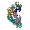
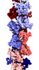
EMDB-34372:
CryoEM structure of the RAD51_ADP filament
EMDB-36040:
Composite map of the Rad51-ADP filament

PDB-8gyk:
CryoEM structure of the RAD51_ADP filament
EMDB-17272:
Structure of RyR1 obtained from SR vesicles tilt series (EMPIAR-10452), reprocessed with TomoBEAR
EMDB-17232:
Purified human apoferritin at 2.8 A resolution from tilt series processed with TomoBEAR

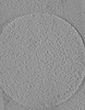
EMDB-16284:
Cryotomogram of L-form-like cytoplasmic membrane vesicles of Listeria monocytogenes Rev2 cell
EMDB-16285:
Cryotomogram of endolysin Ply006 treated Listeria monocytogenes Rev2 cell (stage 1)
ページ:
 wwPDBはEMDBデータモデルのバージョン3へ移行します
