 Movie
Movie Controller

[English] 日本語
 Yorodumi
- EMDB-17272: Structure of RyR1 obtained from SR vesicles tilt series (EMPIAR-1... -
+ Open data

- Basic information
| Entry |  | |||||||||||||||
|---|---|---|---|---|---|---|---|---|---|---|---|---|---|---|---|---|
| Title | Structure of RyR1 obtained from SR vesicles tilt series (EMPIAR-10452), reprocessed with TomoBEAR | |||||||||||||||
 Map data Map data | the nominal apix is 1.8, the calibrated value was found to be 1.7 on that microscope with used mag | |||||||||||||||
 Sample Sample |
| |||||||||||||||
 Keywords Keywords | SR / RyR1 / Ryanodine receptor type 1 / skeletal muscle / sarcoplasmic reticulum / cryo-ET / tomography / StA / subtomogram averaging / tilt series / subnanometer resolution / native membrane / MEMBRANE PROTEIN | |||||||||||||||
| Biological species |  | |||||||||||||||
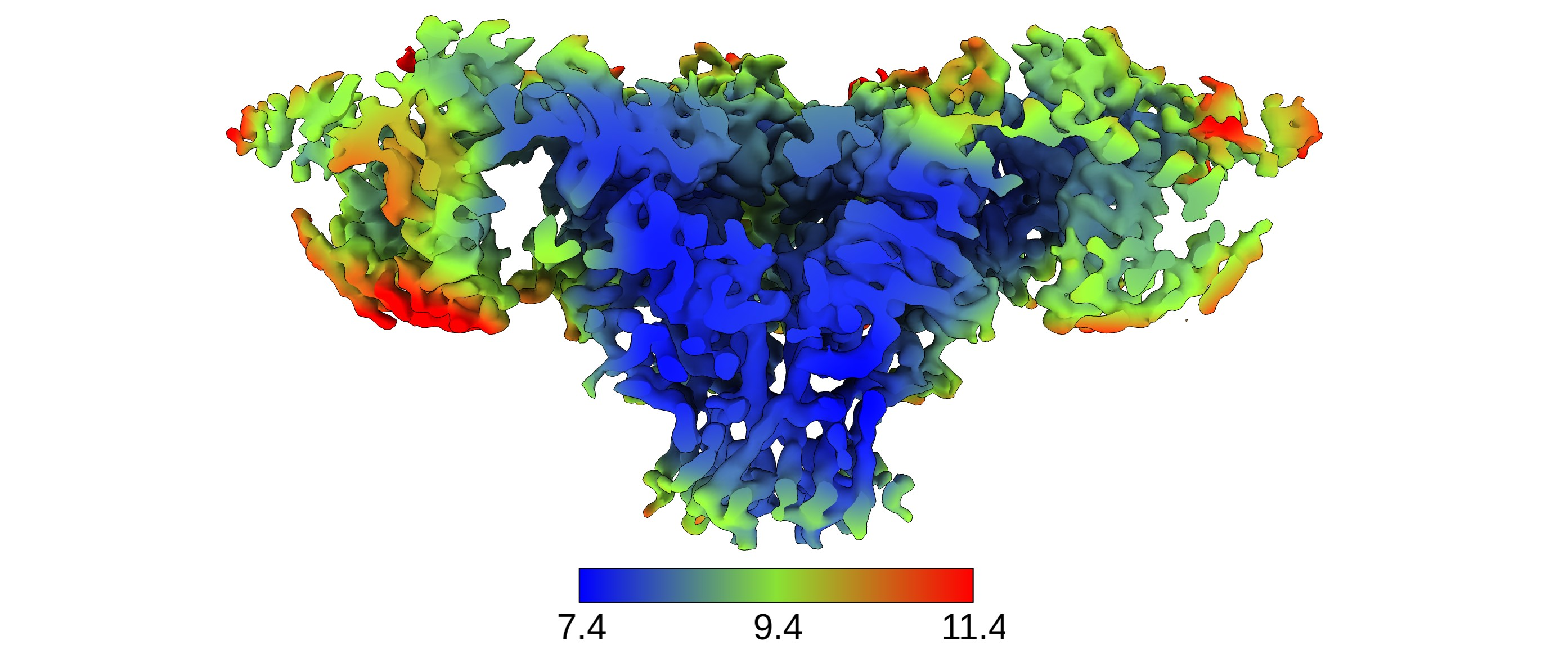
| Method | subtomogram averaging / cryo EM / Resolution: 8.9 Å | |||||||||||||||
 Authors Authors | Balyschew N / Yushkevich A / Mikirtumov V / Sanchez RM / Sprink T / Kudryashev M | |||||||||||||||
| Funding support |  Germany, 4 items Germany, 4 items
| |||||||||||||||
 Citation Citation | Journal: Nat Commun / Year: 2023 Title: Streamlined structure determination by cryo-electron tomography and subtomogram averaging using TomoBEAR. Authors: Nikita Balyschew / Artsemi Yushkevich / Vasilii Mikirtumov / Ricardo M Sanchez / Thiemo Sprink / Mikhail Kudryashev / Abstract: Structures of macromolecules in their native state provide unique unambiguous insights into their functions. Cryo-electron tomography combined with subtomogram averaging demonstrated the power to ...Structures of macromolecules in their native state provide unique unambiguous insights into their functions. Cryo-electron tomography combined with subtomogram averaging demonstrated the power to solve such structures in situ at resolutions in the range of 3 Angstrom for some macromolecules. In order to be applicable to the structural determination of the majority of macromolecules observable in cells in limited amounts, processing of tomographic data has to be performed in a high-throughput manner. Here we present TomoBEAR-a modular configurable workflow engine for streamlined processing of cryo-electron tomographic data for subtomogram averaging. TomoBEAR combines commonly used cryo-EM packages with reasonable presets to provide a transparent ("white box") approach for data management and processing. We demonstrate applications of TomoBEAR to two data sets of purified macromolecular targets, to an ion channel RyR1 in a membrane, and the tomograms of plasma FIB-milled lamellae and demonstrate the ability to produce high-resolution structures. TomoBEAR speeds up data processing, minimizes human interventions, and will help accelerate the adoption of in situ structural biology by cryo-ET. The source code and the documentation are freely available. #1: Journal: bioRxiv / Year: 2023Title: Streamlined Structure Determination by Cryo-Electron Tomography and Subtomogram Averaging using TomoBEAR Authors: Balyschew N / Yushkevich A / Mikirtumov V / Sanchez RM / Sprink T / Kudryashev M | |||||||||||||||
| History |
|
- Structure visualization
| Supplemental images |
|---|
- Downloads & links
-EMDB archive
| Map data | emd_17272.map.gz | 69.7 MB |  EMDB map data format EMDB map data format | |
|---|---|---|---|---|
| Header (meta data) | emd-17272-v30.xmlemd-17272.xml | 16.2 KB 16.2 KB | Display Display | EMDB header |
| FSC (resolution estimation) | emd_17272_fsc.xml | 11.5 KB | Display | FSC data file |
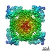
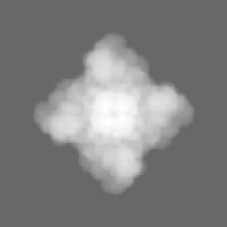
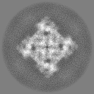
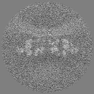
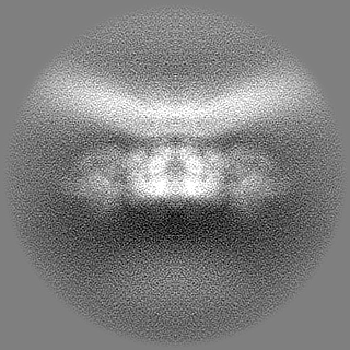
| Images |  emd_17272.png emd_17272.png | 2 MB | ||
| Masks | emd_17272_msk_1.map | 125 MB | Mask map | |
| Filedesc metadata | emd-17272.cif.gz | 4.5 KB | ||
| Others | emd_17272_half_map_1.map.gzemd_17272_half_map_2.map.gz | 61.5 MB 61.5 MB | ||
| Archive directory |  http://ftp.pdbj.org/pub/emdb/structures/EMD-17272ftp://ftp.pdbj.org/pub/emdb/structures/EMD-17272 http://ftp.pdbj.org/pub/emdb/structures/EMD-17272ftp://ftp.pdbj.org/pub/emdb/structures/EMD-17272 | HTTPS FTP |
-Related structure data

-Links
| EMDB pages | EMDB (EBI/PDBe) / EMDataResource |
|---|
-Map
| File | Download / File: emd_17272.map.gz / Format: CCP4 / Size: 125 MB / Type: IMAGE STORED AS FLOATING POINT NUMBER (4 BYTES) | ||||||||||||||||||||||||||||||||||||
|---|---|---|---|---|---|---|---|---|---|---|---|---|---|---|---|---|---|---|---|---|---|---|---|---|---|---|---|---|---|---|---|---|---|---|---|---|---|
| Annotation | the nominal apix is 1.8, the calibrated value was found to be 1.7 on that microscope with used mag | ||||||||||||||||||||||||||||||||||||
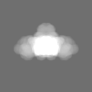
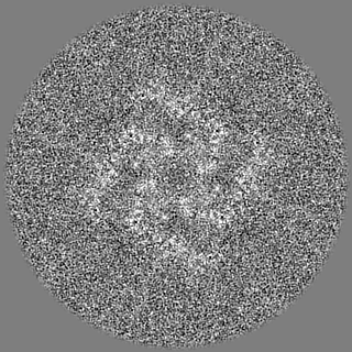
| Projections & slices | Image control
Images are generated by Spider. | ||||||||||||||||||||||||||||||||||||
| Voxel size | X=Y=Z: 1.7 Å | ||||||||||||||||||||||||||||||||||||
| Density |
| ||||||||||||||||||||||||||||||||||||
| Symmetry | Space group: 1 | ||||||||||||||||||||||||||||||||||||
| Details | EMDB XML:
|
 Z (Sec.)
 Y (Row.)
 X (Col.)


-Supplemental data
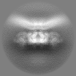
-Mask #1
| File | emd_17272_msk_1.map | ||||||||||||
|---|---|---|---|---|---|---|---|---|---|---|---|---|---|
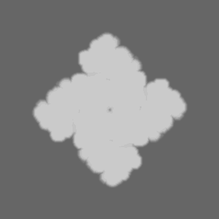
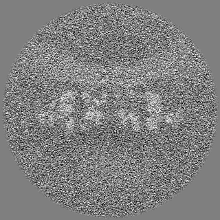
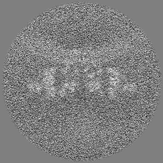
| Projections & Slices |
| ||||||||||||
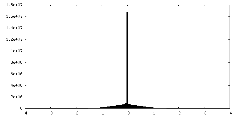
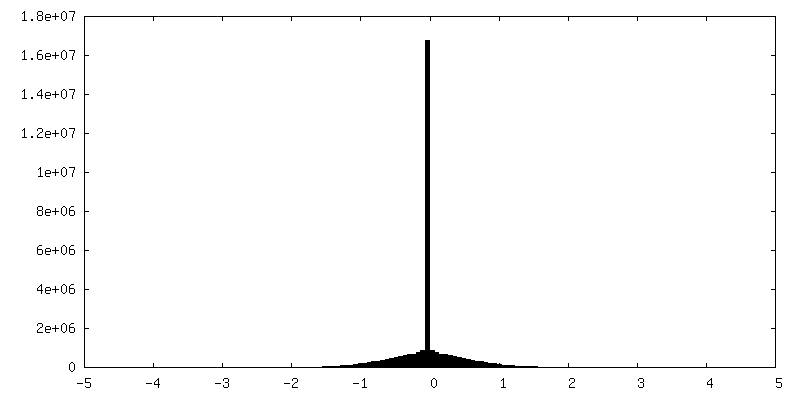
| Density Histograms |


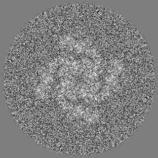
-Half map: #1
| File | emd_17272_half_map_1.map | ||||||||||||
|---|---|---|---|---|---|---|---|---|---|---|---|---|---|
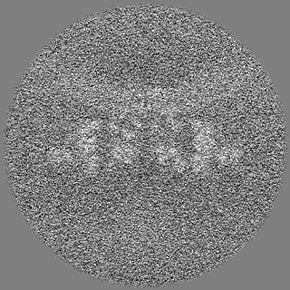
| Projections & Slices |
| ||||||||||||
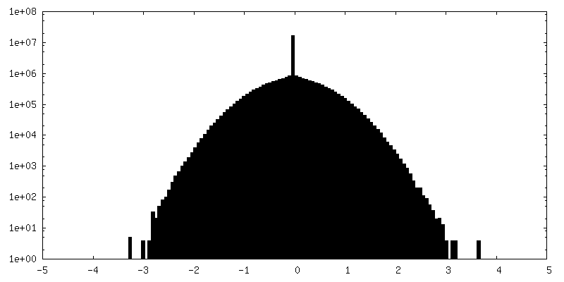
| Density Histograms |

-Half map: #2
| File | emd_17272_half_map_2.map | ||||||||||||
|---|---|---|---|---|---|---|---|---|---|---|---|---|---|
| Projections & Slices |
| ||||||||||||
| Density Histograms |


- Sample components
-Entire : Sarcoplasmic reticulum
| Entire | Name: Sarcoplasmic reticulum |
|---|---|
| Components |
|
-Supramolecule #1: Sarcoplasmic reticulum
| Supramolecule | Name: Sarcoplasmic reticulum / type: organelle_or_cellular_component / ID: 1 / Parent: 0 |
|---|---|
| Source (natural) | Organism: |
-Experimental details
-Structure determination
| Method | cryo EM |
|---|---|
 Processing Processing | subtomogram averaging |
| Aggregation state | tissue |
-Sample preparation
| Buffer | pH: 7.1 |
|---|---|
| Vitrification | Cryogen name: ETHANE |
- Electron microscopy
| Microscope | FEI TITAN KRIOS |
|---|---|
| Image recording | Film or detector model: GATAN K2 SUMMIT (4k x 4k) / Detector mode: COUNTING / Average electron dose: 2.0 e/Å2 |
| Electron beam | Acceleration voltage: 300 kV / Electron source:  FIELD EMISSION GUN FIELD EMISSION GUN |
| Electron optics | Calibrated defocus max: 4.5 µm / Calibrated defocus min: 3.5 µm / Calibrated magnification: 81000 / Illumination mode: FLOOD BEAM / Imaging mode: BRIGHT FIELD / Cs: 2.7 mm / Nominal defocus max: 4.5 µm / Nominal defocus min: 3.5 µm / Nominal magnification: 81000 |
| Sample stage | Specimen holder model: FEI TITAN KRIOS AUTOGRID HOLDER / Cooling holder cryogen: NITROGEN |
| Experimental equipment |  Model: Titan Krios / Image courtesy: FEI Company |
-Image processing
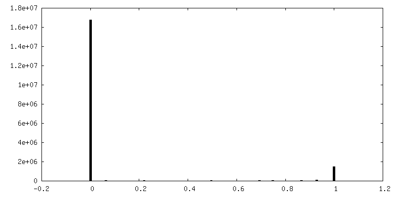
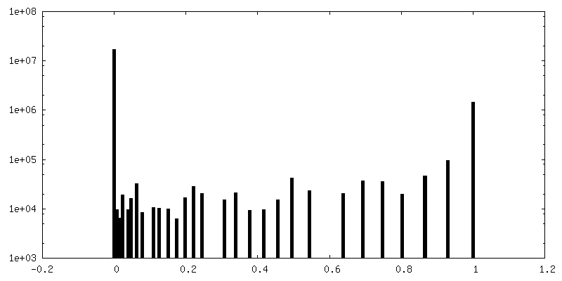
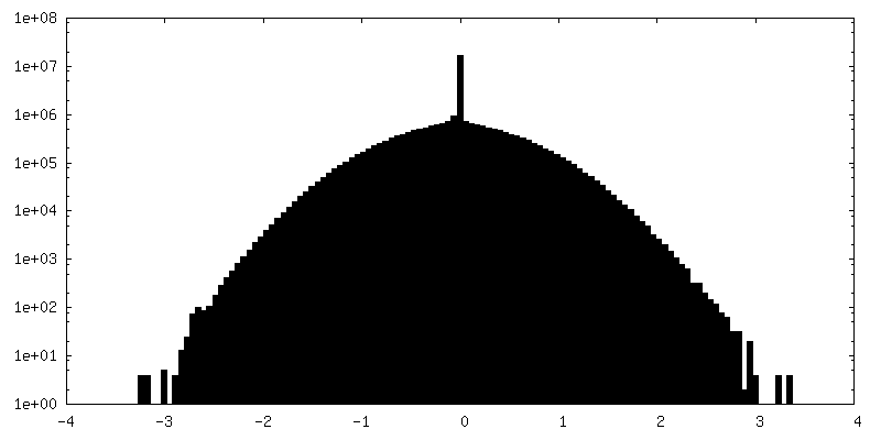
| Final reconstruction | Number classes used: 1 / Applied symmetry - Point group: C4 (4 fold cyclic) / Algorithm: FOURIER SPACE / Resolution.type: BY AUTHOR / Resolution: 8.9 Å / Resolution method: FSC 0.143 CUT-OFF / Software - Name: RELION / Number subtomograms used: 3169 |
|---|---|
| Extraction | Number tomograms: 52 / Number images used: 5200 / Reference model: emd-10840 / Method: template matching / Software - Name: Dynamo / Details: Dynamo GPU template matching in TomoBEAR |
| Final angle assignment | Type: MAXIMUM LIKELIHOOD / Software - Name: RELION (ver. relion 4 beta) |
| FSC plot (resolution estimation) | 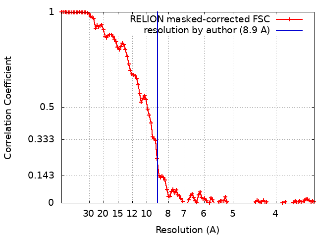 |
