 ムービー
ムービー コントローラー
 構造ビューア
 EMN検索について


-検索条件
-検索結果
検索 (著者・登録者: lai & l)の結果1,975件中、1から50件目までを表示しています
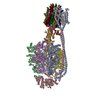
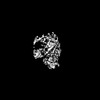
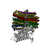
EMDB-50321: 
sub-tomogram averaging map of CHIKV replication organelle connection to the plasma membrane
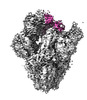
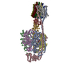
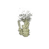
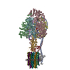
EMDB-39546:
SARS-CoV-2 Delta Spike in complex with JL-8C
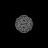
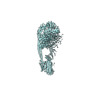
EMDB-39547:
SARS-CoV-2 Delta Spike in complex with JM-1A
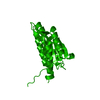
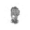
EMDB-39685:
SARS-CoV-2 Delta Spike in complex with Fab of JE-5C
EMDB-39686:
SARS-CoV-2 Spike (BA.1) in complex with Fab of JH-8B

PDB-8yro:
SARS-CoV-2 Delta Spike in complex with JL-8C

PDB-8yrp:
SARS-CoV-2 Delta Spike in complex with JM-1A

PDB-8yz5:
SARS-CoV-2 Delta Spike in complex with Fab of JE-5C

PDB-8yz6:
SARS-CoV-2 Spike (BA.1) in complex with Fab of JH-8B
EMDB-35827:
Structure of CbCas9 bound to 20-nucleotide complementary DNA substrate
EMDB-37652:
Structure of CbCas9 bound to 6-nucleotide complementary DNA substrate
EMDB-37656:
Structure of CbCas9-PcrIIC1 complex bound to 28-bp DNA substrate (20-nt complementary)
EMDB-37657:
Structure of CbCas9-PcrIIC1 complex bound to 62-bp DNA substrate (symmetric 20-nt complementary)
EMDB-37762:
Structure of CbCas9-PcrIIC1 complex bound to 62-bp DNA substrate (non-targeting complex)

PDB-8iyq:
Structure of CbCas9 bound to 20-nucleotide complementary DNA substrate

PDB-8wmh:
Structure of CbCas9 bound to 6-nucleotide complementary DNA substrate

PDB-8wmm:
Structure of CbCas9-PcrIIC1 complex bound to 28-bp DNA substrate (20-nt complementary)

PDB-8wmn:
Structure of CbCas9-PcrIIC1 complex bound to 62-bp DNA substrate (symmetric 20-nt complementary)

PDB-8wr4:
Structure of CbCas9-PcrIIC1 complex bound to 62-bp DNA substrate (non-targeting complex)
PDB-9fhp:
CryoEM structure of wild-type Turnip Yellows Virus

EMDB-40825:
10E8-GT10.2 immunogen in complex with human Fab 10E8 and mouse Fab W6-10
PDB-8sx3:
10E8-GT10.2 immunogen in complex with human Fab 10E8 and mouse Fab W6-10

EMDB-35909:
Cryo-EM structure of Mycobacterium tuberculosis ATP synthase in complex with bedaquiline(BDQ)

EMDB-35911:
Cryo-EM structure of Mycobacterium tuberculosis ATP synthase in the apo-form
PDB-8j0s:
Cryo-EM structure of Mycobacterium tuberculosis ATP synthase in complex with bedaquiline(BDQ)
PDB-8j0t:
Cryo-EM structure of Mycobacterium tuberculosis ATP synthase in the apo-form
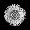
EMDB-50071:
Cryo-EM structure of the icosahedral lumazine synthase from Vicia faba.
PDB-9ez8:
Cryo-EM structure of the icosahedral lumazine synthase from Vicia faba.

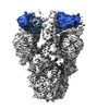
EMDB-17296:
Stabilised BA.1 SARS-CoV-2 spike with H6 nanobodies in '2 up 1 down' RBD conformation

PDB-8oyu:
Stabilised BA.1 SARS-CoV-2 spike with H6 nanobodies in '2 up 1 down' RBD conformation

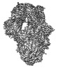
EMDB-19450:
Human UPF1 RNA helicase with AMPPNP
EMDB-19451:
Human UPF1 RNA helicase with AMPPNP and RNA
EMDB-35982:
Cryo-EM structure of Mycobacterium tuberculosis ATP synthase Fo in complex with bedaquiline(BDQ)

EMDB-35983:
Cryo-EM structure of Mycobacterium tuberculosis ATP synthase Fo in the apo-form
EMDB-36015:
Mycobacterium tuberculosis ATP synthase Peripheral Stalk in the apo-form

EMDB-36017:
Mycobacterium tuberculosis ATP synthase Peripheral Stalk in complex with bedaquiline(BDQ)

EMDB-36028:
Mycobacterium tuberculosis ATP synthase F1 in the apo-form
EMDB-36031:
Mycobacterium tuberculosis ATP synthase F1 in complex with bedaquiline(BDQ)
EMDB-36589:
Cryo-EM structure of Mycobacterium tuberculosis ATP synthase in complex with TBAJ-587
EMDB-36590:
Cryo-EM structure of Mycobacterium tuberculosis ATP synthase Fo in complex with TBAJ-587

EMDB-36631:
Cryo-EM structure of Mycobacterium tuberculosis ATP synthase F1 in complex with TBAJ-587
EMDB-36632:
Cryo-EM structure of Mycobacterium tuberculosis ATP synthase Peripheral Stalk in complex with TBAJ-587

EMDB-37243:
Structure of the human ATP synthase bound to bedaquiline (membrane domain)

EMDB-37244:
Structure of the human ATP synthase bound to bedaquiline

EMDB-37245:
Structure of the human ATP synthase bound to bedaquiline (peripheral stalk domain)
EMDB-37251:
Structure of the human ATP synthase bound to bedaquiline (composite)
PDB-8j57:
Cryo-EM structure of Mycobacterium tuberculosis ATP synthase Fo in complex with bedaquiline(BDQ)

PDB-8j58:
Cryo-EM structure of Mycobacterium tuberculosis ATP synthase Fo in the apo-form
PDB-8jr0:
Cryo-EM structure of Mycobacterium tuberculosis ATP synthase in complex with TBAJ-587

PDB-8jr1:
Cryo-EM structure of Mycobacterium tuberculosis ATP synthase Fo in complex with TBAJ-587
ページ:
 wwPDBはEMDBデータモデルのバージョン3へ移行します
