 Movie
Movie Controller


+ Open data

- Basic information
| Entry | Database: PDB / ID: 9fhp | |||||||||
|---|---|---|---|---|---|---|---|---|---|---|
| Title | CryoEM structure of wild-type Turnip Yellows Virus | |||||||||
 Components Components | Minor capsid readthrough protein | |||||||||
 Keywords Keywords | VIRUS / read-through protein | |||||||||
| Function / homology | Potato leaf roll virus readthrough protein / Potato leaf roll virus readthrough protein / Luteovirus group 1 coat protein / Luteovirus coat protein / host cell plasmodesma / host cell periplasmic space / viral capsid / structural molecule activity / Readthrough protein P3-RTD Function and homology information Function and homology information | |||||||||
| Biological species |  Turnip yellows virus Turnip yellows virus | |||||||||
| Method | ELECTRON MICROSCOPY / single particle reconstruction / cryo EM / Resolution: 4.08 Å | |||||||||
 Authors Authors | Trapani, S. / Lai Kee Him, J. / Hoh, F. / Brault, V. / Bron, P. | |||||||||
| Funding support |  France, 1items France, 1items
| |||||||||
 Citation Citation | Journal: Virology / Year: 2025 Title: Structure of the turnip yellows virus particles. Authors: Joséphine Lai-Kee-Him / Stefano Trapani / Sylvaine Boissinot / Catherine Reinbold / Chloé Fallet / Aurélie Ancelin / François Lecorre / François Hoh / Véronique Ziegler-Graff / ...Authors: Joséphine Lai-Kee-Him / Stefano Trapani / Sylvaine Boissinot / Catherine Reinbold / Chloé Fallet / Aurélie Ancelin / François Lecorre / François Hoh / Véronique Ziegler-Graff / Véronique Brault / Patrick Bron / Abstract: Turnip yellows virus (TuYV) is a plant virus infecting important crops such as oilseed rape. TuYV is phloem-restricted and transmitted by aphids. The capsid contains two subunit types: the major ...Turnip yellows virus (TuYV) is a plant virus infecting important crops such as oilseed rape. TuYV is phloem-restricted and transmitted by aphids. The capsid contains two subunit types: the major capsid protein (CP) and a minor component (RTP∗) which arises from the C-terminal cleavage of a readthrough product (RTP). RTP∗ contains the CP sequence fused with a structured domain, denoted RTD, which is a key determinant of virus transmission. Though both CP and RTP∗ are involved in virus movement and aphid transmission, how RTP∗ is incorporated into the capsid is poorly understood. We present here the structural characterisation, by immunogold labelling and 3D cryo-EM, of the wild-type TuYV and a mutant whose capsid contains the CP only. We show that incorporation of RTP∗ does not impair the capsid structure, and the RTD does not adopt well-defined positions at the capsid surface. The number of incorporated RTP∗s suggests a random insertion. | |||||||||
| History |
|
- Structure visualization
| Structure viewer | Molecule: MolmilJmol/JSmol |
|---|
- Downloads & links
-Download
| PDBx/mmCIF format | 9fhp.cif.gz | 85.9 KB | Display | PDBx/mmCIF format |
|---|---|---|---|---|
| PDB format | pdb9fhp.ent.gz | 62.6 KB | Display | PDB format |
| PDBx/mmJSON format | 9fhp.json.gz | Tree view | PDBx/mmJSON format | |
| Others |  Other downloads Other downloads |
-Validation report
| Arichive directory | https://data.pdbj.org/pub/pdb/validation_reports/fh/9fhpftp://data.pdbj.org/pub/pdb/validation_reports/fh/9fhp | HTTPS FTP |
|---|
-Related structure data
| Related structure data | 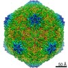 10003MC 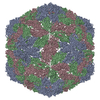 9q8jC M: map data used to model this data C: citing same article ( |
|---|---|
| Similar structure data |
-Links
 PDBj
PDBj- Assembly
| Deposited unit | 
|
|---|---|
| 1 | x 60
|
-Components
| #1: Protein | Mass: 22522.520 Da / Num. of mol.: 3 Source method: isolated from a genetically manipulated source Source: (gene. exp.) Turnip yellows virus / Gene: ORF3/ORF5 / Production host:  Has protein modification | N | |
|---|
-Experimental details
-Experiment
| Experiment | Method: ELECTRON MICROSCOPY |
|---|---|
| EM experiment | Aggregation state: PARTICLE / 3D reconstruction method: single particle reconstruction |
- Sample preparation
| Component | Name: Turnip yellows virus / Type: VIRUS / Details: agroiniltration (English et al. 1997) / Entity ID: all / Source: RECOMBINANT |
|---|---|
| Source (natural) | Organism: Turnip yellows virus |
| Source (recombinant) | Organism: |
| Details of virus | Empty: NO / Enveloped: NO / Isolate: SPECIES / Type: VIRION |
| Buffer solution | pH: 6 |
| Buffer component | Conc.: 0.1 mol/L / Name: citrate |
| Specimen | Conc.: 1.32 mg/ml / Embedding applied: NO / Shadowing applied: NO / Staining applied: NO / Vitrification applied: YES |
| Specimen support | Grid material: COPPER / Grid type: Quantifoil R2/2 |
| Vitrification | Instrument: FEI VITROBOT MARK IV / Cryogen name: ETHANE |
- Electron microscopy imaging
| Experimental equipment |  Model: Tecnai Polara / Image courtesy: FEI Company |
|---|---|
| Microscopy | Model: FEI POLARA 300 |
| Electron gun | Electron source:  FIELD EMISSION GUN / Accelerating voltage: 300 kV / Illumination mode: FLOOD BEAM FIELD EMISSION GUN / Accelerating voltage: 300 kV / Illumination mode: FLOOD BEAM |
| Electron lens | Mode: BRIGHT FIELD / Nominal defocus max: 2500 nm / Nominal defocus min: 800 nm |
| Image recording | Electron dose: 50 e/Å2 / Film or detector model: GATAN K2 QUANTUM (4k x 4k) |
- Processing
| EM software | Name: PHENIX / Version: 1.21.2_5419 / Category: model refinement |
|---|---|
| CTF correction | Type: PHASE FLIPPING AND AMPLITUDE CORRECTION |
| Symmetry | Point symmetry: I (icosahedral) |
| 3D reconstruction | Resolution: 4.08 Å / Resolution method: FSC 0.143 CUT-OFF / Num. of particles: 17316 / Algorithm: FOURIER SPACE / Symmetry type: POINT |
| Atomic model building | Protocol: FLEXIBLE FIT / Space: REAL |
| Atomic model building | PDB-ID: 6rtk 6rtk Accession code: 6rtk / Source name: PDB / Type: experimental model |
| Refinement | Cross valid method: NONE |
