Journal: J Virol / Year: 2024 Title: fate of Chikungunya virus replication organelles. Authors: Justine Girard / Olivier Le Bihan / Joséphine Lai-Kee-Him / Maria Girleanu / Eric Bernard / Cedric Castellarin / Matthew Chee / Aymeric Neyret / Danièle Spehner / Xavier Holy / Anne-Laure ...Authors: Justine Girard / Olivier Le Bihan / Joséphine Lai-Kee-Him / Maria Girleanu / Eric Bernard / Cedric Castellarin / Matthew Chee / Aymeric Neyret / Danièle Spehner / Xavier Holy / Anne-Laure Favier / Laurence Briant / Patrick Bron / Abstract: Chikungunya virus (CHIKV) is a mosquito-borne pathogen responsible for an acute musculoskeletal disease in humans. Replication of the viral RNA genome occurs in specialized membranous replication ...Chikungunya virus (CHIKV) is a mosquito-borne pathogen responsible for an acute musculoskeletal disease in humans. Replication of the viral RNA genome occurs in specialized membranous replication organelles (ROs) or spherules, which contain the viral replication complex. Initially generated by RNA synthesis-associated plasma membrane deformation, alphavirus ROs are generally rapidly endocytosed to produce type I cytopathic vacuoles (CPV-I), from which nascent RNAs are extruded for cytoplasmic translation. By contrast, CHIKV ROs are poorly internalized, raising the question of their fate and functionality at the late stage of infection. Here, using cryogenic-electron microscopy approaches, we investigate the outcome of CHIKV ROs and associated replication machinery in infected human cells. We evidence the late persistence of CHIKV ROs at the plasma membrane with a crowned protein complex at the spherule neck similar to the recently resolved replication complex. The unexpectedly heterogeneous and large diameter of these compartments suggests a continuous, dynamic growth of these organelles beyond the replication of a single RNA genome. Ultrastructural analysis of surrounding cytoplasmic regions supports that outgrown CHIKV ROs remain dynamically active in viral RNA synthesis and export to the cell cytosol for protein translation. Interestingly, rare ROs with a homogeneous diameter are also marginally internalized in CPV-I near honeycomb-like arrangements of unknown function, which are absent in uninfected controls, thereby suggesting a temporal regulation of this internalization. Altogether, this study sheds new light on the dynamic pattern of CHIKV ROs and associated viral replication at the interface with cell membranes in infected cells.IMPORTANCEThe Chikungunya virus (CHIKV) is a positive-stranded RNA virus that requires specialized membranous replication organelles (ROs) for its genome replication. Our knowledge of this viral cycle stage is still incomplete, notably regarding the fate and functional dynamics of CHIKV ROs in infected cells. Here, we show that CHIKV ROs are maintained at the plasma membrane beyond the first viral cycle, continuing to grow and be dynamically active both in viral RNA replication and in its export to the cell cytosol, where translation occurs in proximity to ROs. This contrasts with the homogeneous diameter of ROs during internalization in cytoplasmic vacuoles, which are often associated with honeycomb-like arrangements of unknown function, suggesting a regulated mechanism. This study sheds new light on the dynamics and fate of CHIKV ROs in human cells and, consequently, on our understanding of the Chikungunya viral cycle.
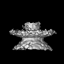
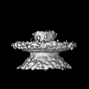
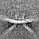
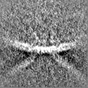
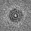
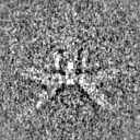
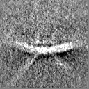
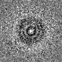
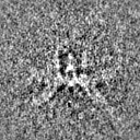
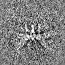
Entire : Chikungunya virus complex at the base of replication organelles
Entire
Name: Chikungunya virus complex at the base of replication organelles
Components
Cell: Chikungunya virus complex at the base of replication organelles
-
Supramolecule #1: Chikungunya virus complex at the base of replication organelles
Supramolecule
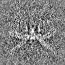
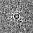
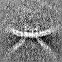
Name: Chikungunya virus complex at the base of replication organelles type: cell / ID: 1 / Parent: 0
Source (natural)
Organism: Chikungunya virus
-
Experimental details
-
Structure determination
Method
cryo EM
Processing
subtomogram averaging
Aggregation state
cell
-
Sample preparation
Buffer
pH: 7.4
Grid
Model: Quantifoil R2/1 / Material: GOLD / Support film - Material: CARBON / Support film - topology: HOLEY / Pretreatment - Type: GLOW DISCHARGE
Vitrification
Cryogen name: ETHANE / Chamber humidity: 100 % / Chamber temperature: 295 K / Instrument: FEI VITROBOT MARK IV Details: 3 microliters of 10 nm BSA-treated fiducial gold particles were added on both sides of the grid before blotting.
Details
HEK293T cells were infected at the MOI of 50 for 17h in the growing medium and then fixed with 4% paraformaldehyde before freezing.
-
Electron microscopy
Microscope
FEI TITAN KRIOS
Image recording
Film or detector model: FEI FALCON III (4k x 4k) / Detector mode: INTEGRATING / Digitization - Dimensions - Width: 4096 pixel / Digitization - Dimensions - Height: 4096 pixel / Average exposure time: 1.3 sec. / Average electron dose: 1.1 e/Å2
Electron beam
Acceleration voltage: 300 kV / Electron source: FIELD EMISSION GUN
In the structure databanks used in Yorodumi, some data are registered as the other names, "COVID-19 virus" and "2019-nCoV". Here are the details of the virus and the list of structure data.
Jan 31, 2019. EMDB accession codes are about to change! (news from PDBe EMDB page)
EMDB accession codes are about to change! (news from PDBe EMDB page)
The allocation of 4 digits for EMDB accession codes will soon come to an end. Whilst these codes will remain in use, new EMDB accession codes will include an additional digit and will expand incrementally as the available range of codes is exhausted. The current 4-digit format prefixed with “EMD-” (i.e. EMD-XXXX) will advance to a 5-digit format (i.e. EMD-XXXXX), and so on. It is currently estimated that the 4-digit codes will be depleted around Spring 2019, at which point the 5-digit format will come into force.
The EM Navigator/Yorodumi systems omit the EMD- prefix.
Related info.:Q: What is EMD? / ID/Accession-code notation in Yorodumi/EM Navigator
Yorodumi is a browser for structure data from EMDB, PDB, SASBDB, etc.
This page is also the successor to EM Navigator detail page, and also detail information page/front-end page for Omokage search.
The word "yorodu" (or yorozu) is an old Japanese word meaning "ten thousand". "mi" (miru) is to see.
Related info.:EMDB / PDB / SASBDB / Comparison of 3 databanks / Yorodumi Search / Aug 31, 2016. New EM Navigator & Yorodumi / Yorodumi Papers / Jmol/JSmol / Function and homology information / Changes in new EM Navigator and Yorodumi
 Movie
Movie Controller

 Yorodumi
 Open data
 Basic information
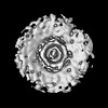
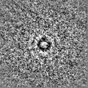
 Map data
 Sample
 Keywords
 Chikungunya virus
 Authors
 France, 1 items
  Citation
 Structure visualization
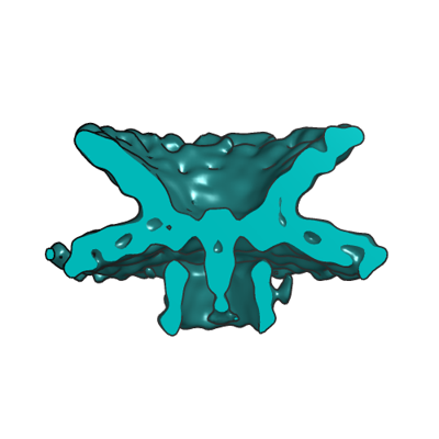
 Downloads & links
 EMDB map data format
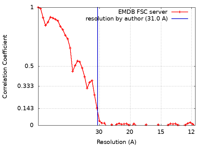
 emd_50321.png
 http://ftp.pdbj.org/pub/emdb/structures/EMD-50321
 Z (Sec.)
 Y (Row.)
 X (Col.)


 Sample components
 Processing
 Electron microscopy
 FIELD EMISSION GUN