 ムービー
ムービー コントローラー
 構造ビューア
 EMN検索について


-検索条件
-検索結果
検索 (著者・登録者: jenkins & j)の結果106件中、1から50件目までを表示しています

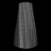
EMDB-44382: 
Structure of the PI4KA complex bound to Calcineurin

PDB-9b9g:
Structure of the PI4KA complex bound to Calcineurin
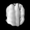
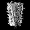
EMDB-18990:
CryoEM map of tau PHF sarkosyl-extracted from a human AD patient (associated with in situ tomography)
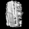
EMDB-50148:
Tau PHF subtomogram average relating to CS1 extended data Figure 9A

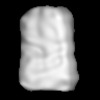
EMDB-50152:
Tau PHF subtomogram average relating to CS2 Figure 3i-j.
EMDB-50153:
Tau PHF subtomogram average relating to CS3 extended data Figure 9c
EMDB-50155:
Tau PHF subtomogram average relating to CS4 extended data Figure 9d

EMDB-50156:
Tau PHF subtomogram average relating to CS5 extended data Figure 9b

EMDB-50157:
Tau PHF subtomogram average relating to CS6 extended data Figure 9e

EMDB-50159:
Tau PHF subtomogram average relating to CS7 extended data Figure 9f
EMDB-50160:
Tau PHF subtomogram average relating to LOL1_PHF Figure 4g-h

EMDB-50161:
Tau SF subtomogram average relating to LOL1_SF Figure 4g-h
EMDB-50162:
Tau SF subtomogram average relating to LOL2_SF Figure 4i-j

EMDB-50358:
In vitro-induced genome-releasing intermediate of Rhodobacter microvirus Ebor computed with C5 symmetry

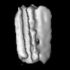
EMDB-50356:
Empty capsid of Rhodobacter microvirus Ebor computed with I4 symmetry

EMDB-50357:
Native capsid of Rhodobacter microvirus Ebor computed with I4 symmetry

EMDB-50359:
Rhodobacter microvirus Ebor attached to B10 host cell reconstructed by single particle analysis with applied C5 symmetry

EMDB-50360:
Rhodobacter microvirus Ebor attached to the outer membrane vesicle

EMDB-50361:
Rhodobacter microvirus Ebor attached to the host cell reconstructed by subtomogram averaging

PDB-9ffg:
Empty capsid of Rhodobacter microvirus Ebor computed with I4 symmetry

PDB-9ffh:
Native capsid of Rhodobacter microvirus Ebor computed with I4 symmetry

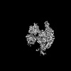
EMDB-40954:
ADP-bound Bcs1 (C7 symmetrized)

EMDB-41061:
ATP-1 state of Bcs1 (C7 symmetrized)

EMDB-41095:
ADP-bound Bcs1 (unsymmetrized)

EMDB-41148:
Apo Bcs1, unsymmetrized

EMDB-41276:
ATP-1 state of Bcs1 (unsymmetrized)

EMDB-41462:
ATP-2 state of Bcs1 (C7 symmetrized)

EMDB-41476:
ATP-2 state of Bcs1 (unsymmetrized)

EMDB-41609:
Bcs1 bound with ISP-ED

PDB-8t14:
ADP-bound Bcs1 (C7 symmetrized)

PDB-8t5u:
ATP-1 state of Bcs1 (C7 symmetrized)

PDB-8t7u:
ADP-bound Bcs1 (unsymmetrized)

PDB-8tby:
Apo Bcs1, unsymmetrized

PDB-8ti0:
ATP-1 state of Bcs1 (unsymmetrized)

PDB-8tp1:
ATP-2 state of Bcs1 (C7 symmetrized)

PDB-8tpl:
ATP-2 state of Bcs1 (unsymmetrized)
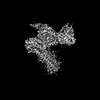
EMDB-19132:
Structure of dynein-2 intermediate chain DYNC2I2 (WDR34) in complex with dynein-2 heavy chain DYNC2H1.

EMDB-19133:
Structure of dynein-2 intermediate chain DYNC2I1 (WDR60) in complex with the dynein-2 heavy chain DYNC2H1.

PDB-8rgg:
Structure of dynein-2 intermediate chain DYNC2I2 (WDR34) in complex with dynein-2 heavy chain DYNC2H1.

PDB-8rgh:
Structure of dynein-2 intermediate chain DYNC2I1 (WDR60) in complex with the dynein-2 heavy chain DYNC2H1.
EMDB-41008:
Cryo-EM structure of the DHA bound FFA4-Gq complex
EMDB-41010:
Cryo-EM structure of the Butyrate bound FFA2-Gq complex
EMDB-41013:
Cryo-EM structure of the DHA bound FFA1-Gq complex

PDB-8t3q:
Cryo-EM structure of the DHA bound FFA4-Gq complex
PDB-8t3s:
Cryo-EM structure of the Butyrate bound FFA2-Gq complex
PDB-8t3v:
Cryo-EM structure of the DHA bound FFA1-Gq complex

EMDB-43013:
Myxococcus xanthus HEnc-K417N(A) protein shell with icosahedral T=1 symmetry

EMDB-43016:
Myxococcus xanthus HEnc-K417N(A) protein shell with tetrahedral symmetry (12 pentamers, 4 hexamers)

EMDB-43037:
Myxococcus xanthus HEnc-K417N(A) protein shell with D3 symmetry (12 pentamers, 3 hexamers)
EMDB-43038:
Myxococcus xanthus HEnc-K417N(A) protein shell with D6 symmetry (12 pentamers, 8 hexamers)
ページ:
 wwPDBはEMDBデータモデルのバージョン3へ移行します
