Movie
Movie Controller


+ Open data

- Basic information
| Entry | Database: PDB / ID: 9b9g | |||||||||||||||||||||||||||||||||||||||
|---|---|---|---|---|---|---|---|---|---|---|---|---|---|---|---|---|---|---|---|---|---|---|---|---|---|---|---|---|---|---|---|---|---|---|---|---|---|---|---|---|
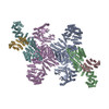
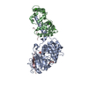
| Title | Structure of the PI4KA complex bound to Calcineurin | |||||||||||||||||||||||||||||||||||||||
 Components Components |
| |||||||||||||||||||||||||||||||||||||||
 Keywords Keywords | SIGNALING PROTEIN / PI4KIIIa complex / PI4KA / TTC7B / FAM126A / CNA / CNB / Calcineurin | |||||||||||||||||||||||||||||||||||||||
| Function / homology |  Function and homology information Function and homology informationreorganization of cellular membranes to establish viral sites of replication / Synthesis of PIPs at the ER membrane / negative regulation of angiotensin-activated signaling pathway / calcium-dependent protein serine/threonine phosphatase regulator activity / regulation of cell proliferation involved in kidney morphogenesis / positive regulation of glomerulus development / negative regulation of calcium ion import across plasma membrane / negative regulation of signaling / calcium-dependent protein serine/threonine phosphatase activity / 1-phosphatidylinositol 4-kinase ...reorganization of cellular membranes to establish viral sites of replication / Synthesis of PIPs at the ER membrane / negative regulation of angiotensin-activated signaling pathway / calcium-dependent protein serine/threonine phosphatase regulator activity / regulation of cell proliferation involved in kidney morphogenesis / positive regulation of glomerulus development / negative regulation of calcium ion import across plasma membrane / negative regulation of signaling / calcium-dependent protein serine/threonine phosphatase activity / 1-phosphatidylinositol 4-kinase / 1-phosphatidylinositol 4-kinase activity / positive regulation of saliva secretion / calmodulin-dependent protein phosphatase activity / calcineurin-NFAT signaling cascade / calcineurin complex / positive regulation of connective tissue replacement / positive regulation of calcium ion-dependent exocytosis of neurotransmitter / positive regulation of calcium ion import across plasma membrane / renal filtration / positive regulation of cardiac muscle hypertrophy in response to stress / host-mediated perturbation of viral process / positive regulation of calcineurin-NFAT signaling cascade / Synthesis of PIPs at the Golgi membrane / Golgi-associated vesicle membrane / skeletal muscle tissue regeneration / phosphatidylinositol biosynthetic process / positive regulation of osteoclast differentiation / dephosphorylation / regulation of synaptic vesicle cycle / positive regulation of activated T cell proliferation / protein dephosphorylation / extrinsic component of plasma membrane / regulation of postsynaptic neurotransmitter receptor internalization / protein-serine/threonine phosphatase / CLEC7A (Dectin-1) induces NFAT activation / epidermis development / phosphatidylinositol phosphate biosynthetic process / positive regulation of osteoblast differentiation / protein serine/threonine phosphatase activity / phosphatidylinositol-mediated signaling / parallel fiber to Purkinje cell synapse / calcineurin-mediated signaling / keratinocyte differentiation / Calcineurin activates NFAT / Activation of BAD and translocation to mitochondria / DARPP-32 events / positive regulation of endocytosis / postsynaptic modulation of chemical synaptic transmission / phosphatase binding / skeletal muscle fiber development / myelination / FCERI mediated Ca+2 mobilization / positive regulation of cell adhesion / protein localization to plasma membrane / hippocampal mossy fiber to CA3 synapse / wound healing / T cell activation / response to calcium ion / sarcolemma / modulation of chemical synaptic transmission / Schaffer collateral - CA1 synapse / Z disc / ATPase binding / Ca2+ pathway / dendritic spine / calmodulin binding / postsynapse / protein dimerization activity / neuron projection / positive regulation of cell migration / cadherin binding / negative regulation of gene expression / protein domain specific binding / focal adhesion / positive regulation of gene expression / calcium ion binding / glutamatergic synapse / enzyme binding / signal transduction / positive regulation of transcription by RNA polymerase II / mitochondrion / extracellular exosome / nucleoplasm / ATP binding / membrane / plasma membrane / cytosol / cytoplasm Similarity search - Function | |||||||||||||||||||||||||||||||||||||||
| Biological species |  Homo sapiens (human) Homo sapiens (human) | |||||||||||||||||||||||||||||||||||||||
| Method | ELECTRON MICROSCOPY / single particle reconstruction / cryo EM / Resolution: 3.5 Å | |||||||||||||||||||||||||||||||||||||||
 Authors Authors | Shaw, A.L. / Suresh, S. / Yip, C.K. / Burke, J.E. | |||||||||||||||||||||||||||||||||||||||
| Funding support |  Canada, 2items Canada, 2items
| |||||||||||||||||||||||||||||||||||||||
 Citation Citation | Journal: Structure / Year: 2024 Title: Structure of calcineurin bound to PI4KA reveals dual interface in both PI4KA and FAM126A. Authors: Alexandria L Shaw / Sushant Suresh / Matthew A H Parson / Noah J Harris / Meredith L Jenkins / Calvin K Yip / John E Burke / Abstract: Phosphatidylinositol 4-kinase alpha (PI4KA) maintains the phosphatidylinositol 4-phosphate (PI4P) and phosphatidylserine pools of the plasma membrane. A key regulator of PI4KA is its association into ...Phosphatidylinositol 4-kinase alpha (PI4KA) maintains the phosphatidylinositol 4-phosphate (PI4P) and phosphatidylserine pools of the plasma membrane. A key regulator of PI4KA is its association into a complex with TTC7 and FAM126 proteins. This complex can be regulated by the CNAβ1 isoform of the phosphatase calcineurin. We previously identified that CNAβ1 directly binds to FAM126A. Here, we report a cryoelectron microscopic (cryo-EM) structure of a truncated PI4KA complex bound to calcineurin, revealing a unique direct interaction between PI4KA and calcineurin. Hydrogen deuterium exchange mass spectrometry (HDX-MS) and computational analysis show that calcineurin forms a complex with an evolutionarily conserved IKISVT sequence in PI4KA's horn domain. We also characterized conserved LTLT and PSISIT calcineurin binding sequences in the C terminus of FAM126A. These dual sites in PI4KA and FAM126A are both in close proximity to phosphorylation sites in the PI4KA complex, suggesting key roles of calcineurin-regulated phosphosites in PI4KA regulation. This work reveals novel insight into how calcineurin can regulate PI4KA activity. | |||||||||||||||||||||||||||||||||||||||
| History |
|
- Structure visualization
| Structure viewer | Molecule: MolmilJmol/JSmol |
|---|
- Downloads & links
-Download
| PDBx/mmCIF format | 9b9g.cif.gz | 1.1 MB | Display | PDBx/mmCIF format |
|---|---|---|---|---|
| PDB format | pdb9b9g.ent.gz | 878.2 KB | Display | PDB format |
| PDBx/mmJSON format | 9b9g.json.gz | Tree view | PDBx/mmJSON format | |
| Others |  Other downloads Other downloads |
-Validation report
| Arichive directory | https://data.pdbj.org/pub/pdb/validation_reports/b9/9b9gftp://data.pdbj.org/pub/pdb/validation_reports/b9/9b9g | HTTPS FTP |
|---|
-Related structure data
| Related structure data |  44382MC M: map data used to model this data C: citing same article ( |
|---|---|
| Similar structure data |
-Links
 PDBj
PDBj


- Assembly
| Deposited unit | 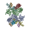
|
|---|---|
| 1 |
|
-Components
-Protein , 5 types, 10 molecules ABDFEGHJIK
| #1: Protein | Mass: 237102.281 Da / Num. of mol.: 2 Source method: isolated from a genetically manipulated source Source: (gene. exp.) Homo sapiens (human) / Gene: PI4KA, PIK4, PIK4CA / Production host:   Spodoptera frugiperda (fall armyworm) Spodoptera frugiperda (fall armyworm)References: UniProt: P42356, 1-phosphatidylinositol 4-kinase #2: Protein | Mass: 94294.109 Da / Num. of mol.: 2 Source method: isolated from a genetically manipulated source Source: (gene. exp.) Homo sapiens (human) / Gene: TTC7B, TTC7L1 / Production host: Spodoptera frugiperda (fall armyworm) / References: UniProt: Q86TV6#3: Protein | Mass: 34638.867 Da / Num. of mol.: 2 Source method: isolated from a genetically manipulated source Details: Truncated construct (1-308) / Source: (gene. exp.) Homo sapiens (human) / Gene: HYCC1, DRCTNNB1A, FAM126A / Production host: Spodoptera frugiperda (fall armyworm) / References: UniProt: Q9BYI3#4: Protein | Mass: 19322.904 Da / Num. of mol.: 2 Source method: isolated from a genetically manipulated source Source: (gene. exp.) Homo sapiens (human) / Gene: PPP3R1, CNA2, CNB / Production host:  #5: Protein | Mass: 70883.242 Da / Num. of mol.: 2 / Mutation: L236P,D238N Source method: isolated from a genetically manipulated source Details: Truncated Calcineurin A alpha (2-391) L236P D238N,Truncated Calcineurin A alpha (2-391) L236P D238N Source: (gene. exp.) Homo sapiens (human) / Gene: PPP3CA, CALNA, CNA / Production host: References: UniProt: Q08209, protein-serine/threonine phosphatase |
|---|
-Non-polymers , 1 types, 8 molecules 
| #6: Chemical | ChemComp-CA /  Mass: 40.078 Da / Num. of mol.: 8 / Source method: obtained synthetically / Formula: Ca Mass: 40.078 Da / Num. of mol.: 8 / Source method: obtained synthetically / Formula: Ca |
|---|
-Details
| Has ligand of interest | N |
|---|---|
| Has protein modification | N |
-Experimental details
-Experiment
| Experiment | Method: ELECTRON MICROSCOPY |
|---|---|
| EM experiment | Aggregation state: PARTICLE / 3D reconstruction method: single particle reconstruction |
- Sample preparation
| Component | Name: PI4KA complex bound to Calcineurin / Type: COMPLEX / Details: Stabilized with BS3 crosslinker / Entity ID: #1-#5 / Source: RECOMBINANT | |||||||||||||||||||||||||
|---|---|---|---|---|---|---|---|---|---|---|---|---|---|---|---|---|---|---|---|---|---|---|---|---|---|---|
| Molecular weight | Experimental value: NO | |||||||||||||||||||||||||
| Source (natural) | Organism: Homo sapiens (human) | |||||||||||||||||||||||||
| Source (recombinant) | Organism: Spodoptera frugiperda (fall armyworm) | |||||||||||||||||||||||||
| Buffer solution | pH: 7 Details: Freshly prepared gel filtration buffer, filtered through 0.22um filter and degassed | |||||||||||||||||||||||||
| Buffer component |
| |||||||||||||||||||||||||
| Specimen | Conc.: 0.77 mg/ml / Embedding applied: NO / Shadowing applied: NO / Staining applied: NO / Vitrification applied: YES Details: Sample was treated with BS3 crosslinker then gel filtered to isolate protein peak consistent with a dimer of pentamers. | |||||||||||||||||||||||||
| Specimen support | Details: Glow discharged using the Pelco EasiGlow. 15mA Current. Grid material: COPPER / Grid mesh size: 300 divisions/in. / Grid type: C-flat-2/1 | |||||||||||||||||||||||||
| Vitrification | Instrument: FEI VITROBOT MARK IV / Cryogen name: ETHANE / Humidity: 100 % / Chamber temperature: 277.15 K / Details: Blot force -5, blot time 1 s |
- Electron microscopy imaging
| Experimental equipment |  Model: Titan Krios / Image courtesy: FEI Company |
|---|---|
| Microscopy | Model: TFS KRIOS |
| Electron gun | Electron source:  FIELD EMISSION GUN / Accelerating voltage: 300 kV / Illumination mode: FLOOD BEAM FIELD EMISSION GUN / Accelerating voltage: 300 kV / Illumination mode: FLOOD BEAM |
| Electron lens | Mode: BRIGHT FIELD / Nominal magnification: 165000 X / Nominal defocus max: 2000 nm / Nominal defocus min: 1000 nm / Cs: 2.7 mm |
| Specimen holder | Cryogen: NITROGEN / Specimen holder model: FEI TITAN KRIOS AUTOGRID HOLDER |
| Image recording | Electron dose: 50 e/Å2 / Film or detector model: FEI FALCON IV (4k x 4k) / Num. of grids imaged: 1 / Num. of real images: 10121 |
| EM imaging optics | Energyfilter name: TFS Selectris |
- Processing
| EM software |
| ||||||||||||||||||||||||||||
|---|---|---|---|---|---|---|---|---|---|---|---|---|---|---|---|---|---|---|---|---|---|---|---|---|---|---|---|---|---|
| CTF correction | Type: PHASE FLIPPING AND AMPLITUDE CORRECTION | ||||||||||||||||||||||||||||
| Particle selection | Num. of particles selected: 1181312 Details: Particles were picked using the cryoSPARC template picker | ||||||||||||||||||||||||||||
| Symmetry | Point symmetry: C2 (2 fold cyclic) | ||||||||||||||||||||||||||||
| 3D reconstruction | Resolution: 3.5 Å / Resolution method: FSC 0.143 CUT-OFF / Num. of particles: 235760 / Symmetry type: POINT | ||||||||||||||||||||||||||||
| Atomic model building |
| ||||||||||||||||||||||||||||
| Refine LS restraints |
|