 ムービー
ムービー コントローラー
 構造ビューア
 EMN検索について


-検索条件
-検索結果
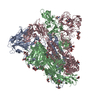
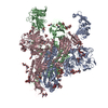
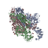
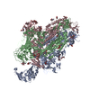
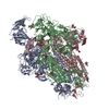
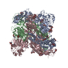
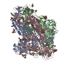
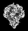
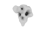
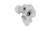
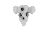
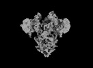
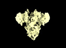
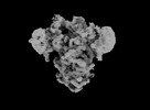
検索 (著者・登録者: draczkowski & p)の結果全42件を表示しています
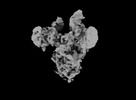
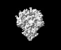
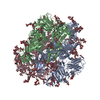
EMDB-33942: 
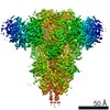
Cryo-EM structure of MERS-CoV spike protein, Two RBD-up conformation 2
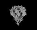
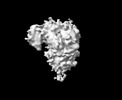
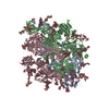
EMDB-33943:
Cryo-EM structure of MERS-CoV spike protein, Two RBD-up conformation 1
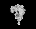
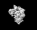
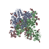
EMDB-33944:
Cryo-EM structure of MERS-CoV spike protein, One RBD-up conformation 4
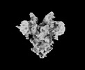
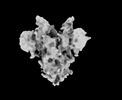
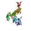
EMDB-33945:
Cryo-EM structure of MERS-CoV spike protein, One RBD-up conformation 3

EMDB-33946:
Cryo-EM structure of MERS-CoV spike protein, One RBD-up conformation 2
EMDB-33947:
Cryo-EM structure of MERS-CoV spike protein, One RBD-up conformation 1
EMDB-33948:
Cryo-EM structure of MERS-CoV spike protein, intermediate conformation
EMDB-33949:
Cryo-EM structure of MERS-CoV spike protein, all RBD-down conformation
PDB-7ymt:
Cryo-EM structure of MERS-CoV spike protein, Two RBD-up conformation 2
PDB-7ymv:
Cryo-EM structure of MERS-CoV spike protein, Two RBD-up conformation 1
PDB-7ymw:
Cryo-EM structure of MERS-CoV spike protein, One RBD-up conformation 4
PDB-7ymx:
Cryo-EM structure of MERS-CoV spike protein, One RBD-up conformation 2
PDB-7ymy:
Cryo-EM structure of MERS-CoV spike protein, One RBD-up conformation 1
PDB-7ymz:
Cryo-EM structure of MERS-CoV spike protein, intermediate conformation
PDB-7yn0:
Cryo-EM structure of MERS-CoV spike protein, all RBD-down conformation
EMDB-32329:
Cryo-EM map of PEDV (Pintung 52) S protein with all three protomers in the D0-down conformation determined in situ on intact viral particles.
EMDB-32332:
Subtomogram averaging of PEDV (Pintung 52) S protein with all three protomers in the D0-down conformation determined in situ on intact viral particles.
EMDB-32333:
Subtomogram averaging of PEDV (Pintung 52) S protein with one protomer in the D0-up conformation and two protomers in the D0-down conformation, determined in situ on intact viral particles
EMDB-32337:
Subtomogram averaging of PEDV (Pintung 52) S protein with two protomers in the D0-up conformation and one protomer in the D0-down conformation, determined in situ on intact viral particles.
EMDB-32338:
Cryo-EM map of PEDV S protein with one protomer in the D0-up conformation while the other two in the D0-down conformation
EMDB-32339:
Subtomogram averaging of PEDV (Pintung 52) S protein with all three protomers in the D0-up conformation determined in situ on intact viral particles.
EMDB-32340:
Subtomogram averaging of PEDV (Pintung 52) S protein in the postfusion form determined in situ on intact viral particles.
EMDB-33646:
Cryo-EM map of IPEC-J2 cell-derived PEDV PT52 S protein with three D0-up
EMDB-33647:
Cryo-EM map of IPEC-J2 cell-derived PEDV PT52 S protein one D0-down and two D0-up
EMDB-33648:
Symmetry-expanded and locally refined protomer structure of IPEC-J2 cell-derived PEDV PT52 S with a CTD-close conformation
EMDB-33649:
Symmetry-expanded and locally refined protomer structure of IPEC-J2 cell-derived PEDV PT52 S with a CTD-open conformation
EMDB-33700:
Cryo-EM map of HEK293F cell-derived PEDV PT52 S protein with three D0-down
EMDB-33701:
Cryo-EM map of HEK293F cell-derived PEDV PT52 S protein one D0-up and two D0-down
EMDB-33702:
Cryo-EM map of HEK293F cell-derived PEDV PT52 S protein with three D0-up
EMDB-33703:
Cryo-EM map of HEK293F cell-derived PEDV PT52 S T326I with three D0-down
EMDB-33704:
Cryo-EM map of HEK293F cell-derived PEDV PT52 S T326I one D0-up and two D0-down
EMDB-33705:
Cryo-EM map of HEK293F cell-derived PEDV PT52 S T326I one D0-down and two D0-up
EMDB-33706:
Cryo-EM map of HEK293F cell-derived PEDV PT52 S T326I with three D0-up
PDB-7w6m:
Cryo-EM map of PEDV (Pintung 52) S protein with all three protomers in the D0-down conformation determined in situ on intact viral particles.
PDB-7w73:
Cryo-EM map of PEDV S protein with one protomer in the D0-up conformation while the other two in the D0-down conformation

PDB-7y6s:
Cryo-EM map of IPEC-J2 cell-derived PEDV PT52 S protein with three D0-up
PDB-7y6t:
Cryo-EM map of IPEC-J2 cell-derived PEDV PT52 S protein one D0-down and two D0-up
PDB-7y6u:
Symmetry-expanded and locally refined protomer structure of IPEC-J2 cell-derived PEDV PT52 S with a CTD-close conformation
PDB-7y6v:
Symmetry-expanded and locally refined protomer structure of IPEC-J2 cell-derived PEDV PT52 S with a CTD-open conformation

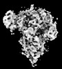
EMDB-30634:
Cryo-EM structure of Ornithine transcarbamylase fused with Ubiquitin in complex with Ubiquitin-carboxy-hydrolase-L1 crosslinked with BS3
EMDB-9891:
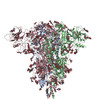
Cryo-EM structure of spike protein of feline infectious peritonitis virus strain UU4
PDB-6jx7:
Cryo-EM structure of spike protein of feline infectious peritonitis virus strain UU4
 wwPDBはEMDBデータモデルのバージョン3へ移行します
