 Movie
Movie Controller
 Structure viewers
 About EMN search


-Search query
-Search result
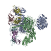
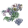
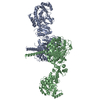
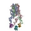
Showing 1 - 50 of 858 items for (author: c. & c. & liu)
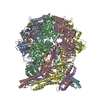
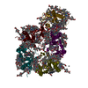
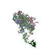
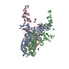
PDB-8kf3: 
The cryo-EM structure of type3 amyloid beta 42 fibril.
PDB-8kf6:
The cryo-EM structure of AV-45 bound type3 amyloid beta 42 fibril.
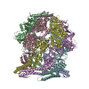
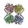
PDB-8kf5:
The cryo-EM structure of type1 amyloid beta 42 fibril in AD3.

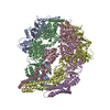
PDB-8kf1:
The cryo-EM structure of AV-45 bound type1 amyloid beta 42 fibril.
PDB-8kf4:
The cryo-EM structure of type1 amyloid beta 42 fibril in AD2 patient.
PDB-8kew:
The cryo-EM structure of type1 amyloid beta 42 fibril.
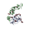
PDB-9atw:
Structure of biofilm-forming functional amyloid PSMa1 from Staphylococcus aureus
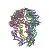
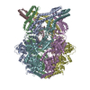
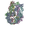
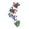
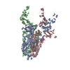
PDB-8k8v:
CryoEM structure of LonC protease hepatmer, apo state
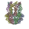
PDB-8k8w:
CryoEM structure of LonC protease open hexamer, apo state
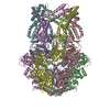
PDB-8k8x:
CryoEM of LonC open pentamer, apo state
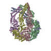
PDB-8k8y:
CryoEM structure of LonC heptamer in presence of AGS
PDB-8k8z:
CryoEM structure of LonC protease hexamer in presence of AGS
PDB-8k90:
CryoEM structure of LonC protease open pentamer in presence of AGS
PDB-8k91:
CryoEM structure of LonC S582A hepatmer with Lysozyme
PDB-8k92:
CryoEM structure of LonC S582A hexamer with Lysozyme
PDB-8k93:
CryoEM structure of LonC protease S582A open hexamer with lysozyme
PDB-8k94:
CryoEM structure of LonC protease S582A open pentamer with lysozyme
PDB-8k95:
CryoEM structure of LonC protease open Hexamer, AGS
PDB-8k96:
CryoEM structure of LonC protease hepatmer with Bortezomib
PDB-8k97:
CryoEM structure of LonC protease hexamer with Bortezomib
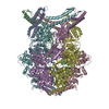
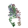
PDB-8tpw:
Crosslinked 6-deoxyerythronolide B synthase (DEBS) Module 3 in complex with antibody fragment 1B2: cis-oriented 1B2 and ACP
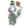
PDB-8tpx:
Crosslinked 6-deoxyerythronolide B synthase (DEBS) Module 3 in complex with antibody fragment 1B2: trans-oriented 1B2 and ACP
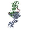
PDB-8wgr:
Cryo-EM structure of inward-open state human norepinephrine transporter NET bound with antidepressant desipramine in KCl condition.
PDB-8wgx:
Cryo-EM structure of inward-open state human norepinephrine transporter NET bound with norepinephrine in nanodisc.

PDB-8z1l:
Cryo-EM structure of human norepinephrine transporter NET in the presence of the antidepressant atomoxetine in an outward-open state at resolution of 3.4 angstrom.
PDB-8wck:
FCP tetramer in Chaetoceros gracilis
PDB-8wcl:
FCP pentamer in Chaetoceros gracilis
PDB-8tko:
KS-AT core of 6-deoxyerythronolide B synthase (DEBS) Module 3 crosslinked with its translocation ACP partner of Module 2
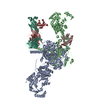
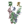
PDB-8vww:
CCHFV GP38 bound to ADI-46152 and ADI-58048 Fabs
PDB-8viw:
Cryo-EM structure of heparosan synthase 2 from Pasteurella multocida with polysaccharide in the GlcNAc-T active site

PDB-8i0i:
dmCTPS with dATP dUTP dGTP and DON
PDB-8tjn:
Crosslinked 6-deoxyerythronolide B synthase (DEBS) Module 1 in complex with antibody fragment 1B2: Crosslinked State 1
PDB-8tjo:
Crosslinked 6-deoxyerythronolide B synthase (DEBS) Module 1 in complex with antibody fragment 1B2: Crosslinked Intra-State 1
PDB-8tjp:
KS-AT core of 6-deoxyerythronolide B synthase (DEBS) Module 3 crosslinked with its elongation ACP partner
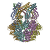
PDB-8y7x:
Structure of HCoV-HKU1A spike in the functionally anchored-3up conformation with 3TMPRSS2
PDB-8y87:
Structure of HCoV-HKU1C spike in the functionally anchored-1up conformation with 1TMPRSS2
PDB-8y88:
Structure of HCoV-HKU1C spike in the functionally anchored-2up conformation with 2TMPRSS2
PDB-8y89:
Structure of HCoV-HKU1C spike in the functionally anchored-3up conformation with 2TMPRSS2
PDB-8y8a:
Structure of HCoV-HKU1C spike in the functionally anchored-3up conformation with 3TMPRSS2
PDB-8y8b:
Local structure of HCoV-HKU1C spike in complex with TMPRSS2 and glycan
PDB-8y8c:
Structure of HCoV-HKU1C spike in the inactive-closed conformation

PDB-8y8d:
Structure of HCoV-HKU1C spike in the inactive-1up conformation
PDB-8y8e:
Structure of HCoV-HKU1C spike in the inactive-2up conformation
PDB-8y8f:
Structure of HCoV-HKU1C spike in the glycan-activated-closed conformation

PDB-8y8g:
Structure of HCoV-HKU1C spike in the glycan-activated-1up conformation
PDB-8y8h:
Structure of HCoV-HKU1C spike in the glycan-activated-2up conformation
PDB-8y8i:
Structure of HCoV-HKU1C spike in the glycan-activated-3up conformation
PDB-8y8j:
Local structure of HCoV-HKU1C spike in complex with glycan
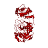
PDB-8vd7:
MicroED structure of SARS-CoV-2 main protease (MPro/3CLPro) with missing cone eliminated by suspended drop
PDB-9ijp:
Polyphosphate bound alpha-synuclein fibril
Pages:
 wwPDB to switch to version 3 of the EMDB data model
