 Movie
Movie Controller

[English] 日本語
 Yorodumi
- PDB-8viw: Cryo-EM structure of heparosan synthase 2 from Pasteurella multoc... -
+ Open data

- Basic information
| Entry | Database: PDB / ID: 8viw | ||||||
|---|---|---|---|---|---|---|---|
| Title | Cryo-EM structure of heparosan synthase 2 from Pasteurella multocida with polysaccharide in the GlcNAc-T active site | ||||||
 Components Components | Heparosan synthase B | ||||||
 Keywords Keywords | TRANSFERASE / polysaccharide synthase / complex | ||||||
| Function / homology | Glycosyltransferase 2-like / Glycosyl transferase family 2 / hexosyltransferase activity / Nucleotide-diphospho-sugar transferases / nucleotide binding / metal ion binding / : / URIDINE-5'-DIPHOSPHATE / Heparosan synthase B Function and homology information Function and homology information | ||||||
| Biological species |  Pasteurella multocida (bacteria) Pasteurella multocida (bacteria) | ||||||
| Method | ELECTRON MICROSCOPY / single particle reconstruction / cryo EM / Resolution: 3.3 Å | ||||||
 Authors Authors | Krahn, J.M. / Pedersen, L.C. / Liu, J. / Stancanelli, E. / Borgnia, M. / Vivarette, E. | ||||||
| Funding support |  United States, 1items United States, 1items
| ||||||
 Citation Citation | Journal: ACS Catal / Year: 2024 Title: Structural and Functional Analysis of Heparosan Synthase 2 from (PmHS2) to Improve the Synthesis of Heparin. Authors: Eduardo Stancanelli / Juno A Krahn / Elizabeth Viverette / Robert Dutcher / Vijayakanth Pagadala / Mario J Borgnia / Jian Liu / Lars C Pedersen / Abstract: Heparin is a widely used drug to treat thrombotic disorders in hospitals. Heparosan synthase 2 from (PmHS2) is a key enzyme used for the chemoenzymatic synthesis of heparin oligosaccharides. It has ...Heparin is a widely used drug to treat thrombotic disorders in hospitals. Heparosan synthase 2 from (PmHS2) is a key enzyme used for the chemoenzymatic synthesis of heparin oligosaccharides. It has both activities: glucosaminyl transferase activity and glucuronyl transferase activity; however, the mechanism to carry out the glyco-oligomerization is unknown. Here, we report crystal structures of PmHS2 constructs with bound uridine diphosphate (UDP) and a cryo-EM structure of PmHS2 in complex with UDP and a heptasaccharide (NS 7-mer) substrate. Using a LC-MC analytical method, we discovered the enzyme displays both a two-step concerted oligomerization mode and a distributive oligomerization mode depending on the non-reducing end of the starting oligosaccharide primer. Removal of 7 amino acid residues from the C-terminus results in an enzymatically active monomer instead of dimer and loses the concerted oligomerization mode of activity. In addition, the monomer construct can transfer N-acetyl glucosamine at a substrate concentration that is ∼7-fold higher than wildtype enzyme. It was also determined that an F529A mutant can transfer an N-sulfo glucosamine (GlcNS) saccharide from a previously inactive UDP-GlcNS donor. Performing the glyco-transfer reaction at a high substrate concentration and the capability of using unnatural donors are desirable to simplify the chemoenzymatic synthesis to prepare heparin-based therapeutics. | ||||||
| History |
|
- Structure visualization
| Structure viewer | Molecule: MolmilJmol/JSmol |
|---|
- Downloads & links
-Download
| PDBx/mmCIF format | 8viw.cif.gz | 405.5 KB | Display | PDBx/mmCIF format |
|---|---|---|---|---|
| PDB format | pdb8viw.ent.gz | 326.9 KB | Display | PDB format |
| PDBx/mmJSON format | 8viw.json.gz | Tree view | PDBx/mmJSON format | |
| Others |  Other downloads Other downloads |
-Validation report
| Arichive directory | https://data.pdbj.org/pub/pdb/validation_reports/vi/8viwftp://data.pdbj.org/pub/pdb/validation_reports/vi/8viw | HTTPS FTP |
|---|
-Related structure data
| Related structure data |  43269MC 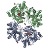 8vh7C 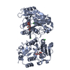 8vh8C C: citing same article ( M: map data used to model this data |
|---|---|
| Similar structure data |
-Links
 PDBj
PDBj


- Assembly
| Deposited unit | 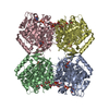
|
|---|---|
| 1 |
|
-Components
| #1: Protein | Mass: 64548.277 Da / Num. of mol.: 4 Source method: isolated from a genetically manipulated source Source: (gene. exp.) Pasteurella multocida (bacteria) / Gene: hssB / Production host: #2: Polysaccharide | beta-D-glucopyranuronic acid-(1-4)-2-deoxy-2-(sulfoamino)-alpha-D-glucopyranose-(1-4)-beta-D- ...beta-D-glucopyranuronic acid-(1-4)-2-deoxy-2-(sulfoamino)-alpha-D-glucopyranose-(1-4)-beta-D-glucopyranuronic acid-(1-4)-2-deoxy-2-(sulfoamino)-alpha-D-glucopyranose-(1-4)-beta-D-glucopyranuronic acid Type: oligosaccharide / Mass: 1028.824 Da / Num. of mol.: 4 Source method: isolated from a genetically manipulated source #3: Chemical | ChemComp-MN /   Mass: 54.938 Da / Num. of mol.: 8 / Source method: obtained synthetically / Formula: Mn / Feature type: SUBJECT OF INVESTIGATION Mass: 54.938 Da / Num. of mol.: 8 / Source method: obtained synthetically / Formula: Mn / Feature type: SUBJECT OF INVESTIGATION#4: Chemical | ChemComp-UDP /   Type: RNA linking / Mass: 404.161 Da / Num. of mol.: 8 / Source method: obtained synthetically / Formula: C9H14N2O12P2 / Feature type: SUBJECT OF INVESTIGATION / Comment: UDP*YM Type: RNA linking / Mass: 404.161 Da / Num. of mol.: 8 / Source method: obtained synthetically / Formula: C9H14N2O12P2 / Feature type: SUBJECT OF INVESTIGATION / Comment: UDP*YM#5: Water | ChemComp-HOH / |  Mass: 18.015 Da / Num. of mol.: 4 / Source method: isolated from a natural source / Formula: H2O Mass: 18.015 Da / Num. of mol.: 4 / Source method: isolated from a natural source / Formula: H2OHas ligand of interest | Y | Has protein modification | N | |
|---|
-Experimental details
-Experiment
| Experiment | Method: ELECTRON MICROSCOPY |
|---|---|
| EM experiment | Aggregation state: 2D ARRAY / 3D reconstruction method: single particle reconstruction |
- Sample preparation
| Component | Name: pmHS2 with 7-mer polysaccharide / Type: ORGANELLE OR CELLULAR COMPONENT / Entity ID: #1 / Source: RECOMBINANT |
|---|---|
| Molecular weight | Experimental value: NO |
| Source (natural) | Organism: Pasteurella multocida (bacteria) |
| Source (recombinant) | Organism: |
| Buffer solution | pH: 7.5 Details: 25 mM Tris pH 7.5, 87.5 mM NaCl, 1 mM MnCl2, 1 mM UDP and 1 mM NS-7mer (GlcA-GlcNS-GlcA-GlcNS-GlcA-GlcNS-GlcA-pNP) |
| Specimen | Conc.: 0.5 mg/ml / Embedding applied: NO / Shadowing applied: NO / Staining applied: NO / Vitrification applied: YES |
| Specimen support | Grid material: GOLD / Grid type: UltrAuFoil R1.2/1.3 |
| Vitrification | Cryogen name: NITROGEN |
- Electron microscopy imaging
| Experimental equipment |  Model: Titan Krios / Image courtesy: FEI Company |
|---|---|
| Microscopy | Model: FEI TITAN KRIOS |
| Electron gun | Electron source:  FIELD EMISSION GUN / Accelerating voltage: 300 kV / Illumination mode: SPOT SCAN FIELD EMISSION GUN / Accelerating voltage: 300 kV / Illumination mode: SPOT SCAN |
| Electron lens | Mode: BRIGHT FIELD / Nominal magnification: 130000 X / Nominal defocus max: 1800 nm / Nominal defocus min: 1000 nm / Cs: 2.7 mm |
| Image recording | Electron dose: 50 e/Å2 / Film or detector model: GATAN K3 (6k x 4k) / Num. of real images: 4544 |
- Processing
| EM software |
| ||||||||||||
|---|---|---|---|---|---|---|---|---|---|---|---|---|---|
| CTF correction | Type: PHASE FLIPPING AND AMPLITUDE CORRECTION | ||||||||||||
| 3D reconstruction | Resolution: 3.3 Å / Resolution method: FSC 0.143 CUT-OFF / Num. of particles: 104255 / Algorithm: FOURIER SPACE / Num. of class averages: 169 / Symmetry type: POINT | ||||||||||||
| Refinement | Cross valid method: NONE |
