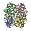
[English] 日本語
Yorodumi
- PDB-8vd7: MicroED structure of SARS-CoV-2 main protease (MPro/3CLPro) with ... -

+
Open data


ID or keywords:

Loading...

-
Basic information

Entry
Database: PDB / ID: 8vd7
TitleMicroED structure of SARS-CoV-2 main protease (MPro/3CLPro) with missing cone eliminated by suspended drop
Components3C-like proteinase nsp5
KeywordsVIRAL PROTEIN / Cysteine Protease / Viral Protease / Viral Assembly
Function / homology
Function and homology information


protein guanylyltransferase activity / RNA endonuclease activity, producing 3'-phosphomonoesters / mRNA guanylyltransferase activity / 5'-3' RNA helicase activity / Lyases; Phosphorus-oxygen lyases / symbiont-mediated suppression of host cytoplasmic pattern recognition receptor signaling pathway via inhibition of TBK1 activity / Assembly of the SARS-CoV-2 Replication-Transcription Complex (RTC) / Maturation of replicase proteins / ISG15-specific peptidase activity / Transcription of SARS-CoV-2 sgRNAs ...protein guanylyltransferase activity / RNA endonuclease activity, producing 3'-phosphomonoesters / mRNA guanylyltransferase activity / 5'-3' RNA helicase activity / Lyases; Phosphorus-oxygen lyases / symbiont-mediated suppression of host cytoplasmic pattern recognition receptor signaling pathway via inhibition of TBK1 activity / Assembly of the SARS-CoV-2 Replication-Transcription Complex (RTC) / Maturation of replicase proteins / ISG15-specific peptidase activity / Transcription of SARS-CoV-2 sgRNAs / Translation of Replicase and Assembly of the Replication Transcription Complex / TRAF3-dependent IRF activation pathway / Replication of the SARS-CoV-2 genome / snRNP Assembly / double membrane vesicle viral factory outer membrane / Hydrolases; Acting on ester bonds; Exoribonucleases producing 5'-phosphomonoesters / 5'-3' DNA helicase activity / SARS coronavirus main proteinase / host cell endoplasmic reticulum-Golgi intermediate compartment / 3'-5'-RNA exonuclease activity / host cell endosome / symbiont-mediated suppression of host toll-like receptor signaling pathway / symbiont-mediated degradation of host mRNA / mRNA guanylyltransferase / symbiont-mediated suppression of host ISG15-protein conjugation / G-quadruplex RNA binding / omega peptidase activity / symbiont-mediated suppression of host cytoplasmic pattern recognition receptor signaling pathway via inhibition of IRF3 activity / SARS-CoV-2 modulates host translation machinery / mRNA (guanine-N7)-methyltransferase / host cell Golgi apparatus / methyltransferase cap1 / symbiont-mediated perturbation of host ubiquitin-like protein modification / DNA helicase / mRNA (nucleoside-2'-O-)-methyltransferase activity / mRNA 5'-cap (guanine-N7-)-methyltransferase activity / ubiquitinyl hydrolase 1 / cysteine-type deubiquitinase activity / Hydrolases; Acting on peptide bonds (peptidases); Cysteine endopeptidases / single-stranded RNA binding / host cell perinuclear region of cytoplasm / host cell endoplasmic reticulum membrane / viral protein processing / lyase activity / symbiont-mediated suppression of host type I interferon-mediated signaling pathway / RNA helicase / induction by virus of host autophagy / copper ion binding / RNA-directed RNA polymerase / viral RNA genome replication / cysteine-type endopeptidase activity / RNA-dependent RNA polymerase activity / virus-mediated perturbation of host defense response / DNA-templated transcription / lipid binding / host cell nucleus / SARS-CoV-2 activates/modulates innate and adaptive immune responses / ATP hydrolysis activity / proteolysis / RNA binding / zinc ion binding / ATP binding / membrane
Similarity search - Function
RNA-dependent RNA polymerase, SARS-CoV-like / : / Nonstructural protein 14, betacoronavirus / NSP15, NendoU domain, coronavirus / : / : / Coronavirus Nonstructural protein 13, 1B domain / Coronavirus Non-structural protein 13, zinc-binding domain / Coronavirus Nonstructural protein 13, stalk domain / : ...RNA-dependent RNA polymerase, SARS-CoV-like / : / Nonstructural protein 14, betacoronavirus / NSP15, NendoU domain, coronavirus / : / : / Coronavirus Nonstructural protein 13, 1B domain / Coronavirus Non-structural protein 13, zinc-binding domain / Coronavirus Nonstructural protein 13, stalk domain / : / Coronavirus Nsp12 Interface domain profile. / Nonstructural protein 15, middle domain, alpha/betacoronavirus / Nonstructural protein 15, N-terminal domain, alpha/beta-coronavirus / NSP14, guanine-N7-methyltransferase domain, coronavirus / NSP12 RNA-dependent RNA polymerase, coronavirus / Coronavirus (CoV) guanine-N7-methyltransferase (N7-MTase) domain profile. / Coronavirus (CoV) Nsp15 N-terminal oligomerization domain profile. / Nidovirus 2-O-methyltransferase / Coronavirus Nsp12 RNA-dependent RNA polymerase (RdRp) domain profile. / Nidovirus 3'-5' exoribonuclease domain / Nidovirus 2'-O-methyltransferase (2'-O-MTase) domain profile. / Nidovirus 3'-5' exoribonuclease (ExoN) domain profile. / Nonstructural protein 13, 1B domain, coronavirus / Arterivirus Nsp11 N-terminal/Coronavirus NSP15 middle domain / Non-structural protein NSP15, N-terminal domain superfamily, coronavirus / Non-structural protein NSP15, middle domain superfamily / Coronavirus replicase NSP15, N-terminal oligomerization / Nonstructural protein 15, middle domain, coronavirus / Coronavirus replicase NSP15, middle domain / Coronavirus replicase NSP15, N-terminal oligomerisation / Arterivirus Nsp11 N-terminal/coronavirus NSP15 middle (AV-Nsp11N/CoV-Nsp15M) domain profile. / Non-structural protein NSP16, coronavirus-like / Non-structural protein 14, coronavirus / RNA polymerase, N-terminal, coronavirus / Coronavirus 2'-O-methyltransferase / Coronavirus proofreading exoribonuclease / Coronavirus RNA-dependent RNA polymerase, N-terminal / Nidoviral uridylate-specific endoribonuclease (NendoU) domain profile. / Nidovirus RdRp-associated nucleotidyl transferase (NiRAN) domain / Nonstructural protein 13, zinc-binding domain, coronavirus-like / Coronaviridae zinc-binding (CV ZBD) domain profile. / Nidovirus RdRp-associated nucleotidyl transferase (NiRAN) domain profile. / Endoribonuclease EndoU-like / NendoU domain, nidovirus / Coronavirus replicase NSP15, uridylate-specific endoribonuclease / DNA2/NAM7 helicase-like, C-terminal / AAA domain / Lipocalin signature. / (+) RNA virus helicase core domain / (+)RNA virus helicase core domain profile. / Non-structural protein NSP3, SUD-N (Mac2) domain, betacoronavirus / Sarbecovirus Nsp3c-N domain profile. / Non-structural protein NSP3, N-terminal, betacoronavirus / Polyprotein cleavage domain PL2pro superfamily, betacoronavirus / Non-structural protein NSP3, SUD-N (Mac2) domain superfamily, betacoronavirus / Betacoronavirus SUD-C domain / Betacoronavirus replicase NSP3, N-terminal / NSP1 globular domain superfamily, betacoronavirus / Non-structural protein 2, SARS-CoV-like / Carbamoyl-phosphate synthase subdomain signature 2. / : / Betacoronavirus Nsp3e group 2-specific marker (G2M) domain profile. / NSP1, C-terminal domain, betacoronavirus / Betacoronavirus Nsp3c-M domain profile. / NSP1, globular domain, betacoronavirus / Non-structural protein NSP3, SUD-M domain, betacoronavirus / Non-structural protein NSP3, SUD-M domain superfamily, betacoronavirus / Betacoronavirus replicase NSP1 / Betacoronavirus single-stranded poly(A) binding domain / Betacoronavirus (BetaCoV) Nsp1 C-terminal domain profile. / Betacoronavirus Nsp3c-C domain profile. / Betacoronavirus Nsp3e nucleic acid-binding (NAB) domain profile. / DPUP/SUD, C-terminal, betacoronavirus / Non-structural protein NSP3, nucleic acid-binding domain superfamily, betacoronavirus / Non-structural protein 6, betacoronavirus / Betacoronavirus nucleic acid-binding (NAB) / Non-structural protein NSP3, nucleic acid-binding domain, betacoronavirus / Non-structural protein NSP3A domain-like superfamily / Papain-like protease, N-terminal domain superfamily, coronavirus / Papain-like viral protease, palm and finger domains, coronavirus / : / Coronavirus 3Ecto domain profile. / : / Coronavirus (CoV) Nsp2 middle domain profile. / Coronavirus (CoV) Nsp2 N-terminal domain profile. / Coronavirus (CoV) Nsp2 C-terminal domain profile. / NSP1, globular domain, alpha/betacoronavirus / : / Coronavirus (CoV) Nsp3 Y domain profile. / Coronavirus (CoV) Nsp1 globular domain profile. / Coronavirus replicase NSP2, N-terminal / Nonstructural protein 2, N-terminal domain, coronavirus / Coronavirus replicase NSP2, C-terminal / Non-structural protein 2, C-terminal domain, coronavirus / Coronavirus Nsp3a Ubl domain profile. / Coronavirus Nsp3d Ubl domain profile. / Coronavirus RNA-dependent RNA polymerase (RdRp) Nsp7 cofactor domain profile. / Coronavirus RNA-dependent RNA polymerase (RdRp) Nsp8 cofactor domain profile. / Coronavirus Nsp9 single-stranded RNA (ssRNA)-binding domain profile. / Coronavirus (CoV) ExoN/MTase coactivator domain profile.
Similarity search - Domain/homology
Replicase polyprotein 1ab
Similarity search - Component
Biological speciesSevere acute respiratory syndrome coronavirus 2
MethodELECTRON CRYSTALLOGRAPHY / electron crystallography / MOLECULAR REPLACEMENT / cryo EM / Resolution: 2.15 Å
AuthorsBu, G. / Gillman, C. / Danelius, E. / Hattne, J. / Nannenga, B.L. / Gonen, T.
Funding support United States, 3items
OrganizationGrant numberCountry
National Institutes of Health/National Institute of General Medical Sciences (NIH/NIGMS)P41GM136508 United States
Department of Defense (DOD, United States)HDTRA1-21-1-0004 United States
Howard Hughes Medical Institute (HHMI) United States
CitationJournal: J Struct Biol X / Year: 2024
Title: Eliminating the missing cone challenge through innovative approaches.
Authors: Cody Gillman / Guanhong Bu / Emma Danelius / Johan Hattne / Brent L Nannenga / Tamir Gonen /
Abstract: Microcrystal electron diffraction (MicroED) has emerged as a powerful technique for unraveling molecular structures from microcrystals too small for X-ray diffraction. However, a significant hurdle ...Microcrystal electron diffraction (MicroED) has emerged as a powerful technique for unraveling molecular structures from microcrystals too small for X-ray diffraction. However, a significant hurdle arises with plate-like crystals that consistently orient themselves flat on the electron microscopy grid. If the normal of the plate correlates with the axes of the crystal lattice, the crystal orientations accessible for measurement are restricted because the crystal cannot be arbitrarily rotated. This limits the information that can be acquired, resulting in a missing cone of information. We recently introduced a novel crystallization strategy called suspended drop crystallization and proposed that crystals in a suspended drop could effectively address the challenge of preferred crystal orientation. Here we demonstrate the success of the suspended drop approach in eliminating the missing cone in two samples that crystallize as thin plates: bovine liver catalase and the SARS‑CoV‑2 main protease (Mpro). This innovative solution proves indispensable for crystals exhibiting systematic preferred orientations, unlocking new possibilities for structure determination by MicroED.
History
DepositionDec 14, 2023Deposition site: RCSB / Processing site: RCSB
Revision 1.0Jul 17, 2024Provider: repository / Type: Initial release

-
Structure visualization

Structure viewerMolecule:
MolmilJmol/JSmol

Downloads & links

-
Assembly

Deposited unit
A: 3C-like proteinase nsp5
hetero molecules


Theoretical massNumber of molelcules
Total (without water)33,8612
Polymers33,8261
Non-polymers351
Water1086
1
A: 3C-like proteinase nsp5
hetero molecules

A: 3C-like proteinase nsp5
hetero molecules


Theoretical massNumber of molelcules
Total (without water)67,7224
Polymers67,6512
Non-polymers712
Water362
TypeNameSymmetry operationNumber
identity operation1_555x,y,z1
point symmetry operation1
Buried area3420 Å2
ΔGint-31 kcal/mol
Surface area26440 Å2
MethodPISA
Unit cell
Length a, b, c (Å)115.540, 55.530, 45.370
Angle α, β, γ (deg.)90.000, 101.074, 90.000
Int Tables number5
Space group name H-MC121
Symmetry operation#1: x,y,z
#2: -x,y,-z
#3: x+1/2,y+1/2,z
#4: -x+1/2,y+1/2,-z
Components on special symmetry positions
IDModelComponents
11A-501-

CL

-
Components

#1: Protein 3C-like proteinase nsp5 / 3CL-PRO / 3CLp / Main protease / Mpro / Non-structural protein 5 / nsp5 / SARS coronavirus main proteinase


Mass: 33825.547 Da / Num. of mol.: 1
Source method: isolated from a genetically manipulated source
Source: (gene. exp.) Severe acute respiratory syndrome coronavirus 2
Gene: rep, 1a-1b / Production host: Escherichia coli (E. coli)
References: UniProt: P0DTD1, SARS coronavirus main proteinase
#2: Chemical ChemComp-CL / CHLORIDE ION


Mass: 35.453 Da / Num. of mol.: 1 / Source method: obtained synthetically / Formula: Cl
#3: Water ChemComp-HOH / water


Mass: 18.015 Da / Num. of mol.: 6 / Source method: isolated from a natural source / Formula: H2O
Has ligand of interestN

-
Experimental details

-
Experiment

ExperimentMethod: ELECTRON CRYSTALLOGRAPHY
EM experimentAggregation state: 3D ARRAY / 3D reconstruction method: electron crystallography

-
Sample preparation

ComponentName: SARS-CoV-2 main protease (Mpro) / Type: COMPLEX / Entity ID: #1 / Source: RECOMBINANT
Source (natural)Organism: Severe acute respiratory syndrome coronavirus 2
Source (recombinant)Organism: Escherichia coli (E. coli)
Buffer solutionpH: 6.5 / Details: 0.1 M MES pH 6.5, 20% PEG 3350, 5% DMSO.
SpecimenEmbedding applied: NO / Shadowing applied: NO / Staining applied: NO / Vitrification applied: YES
VitrificationCryogen name: ETHANE / Humidity: 95 % / Chamber temperature: 293 K

-
Data collection

Experimental equipment
Model: Titan Krios / Image courtesy: FEI Company
MicroscopyModel: FEI TITAN KRIOS
Electron gunElectron source: FIELD EMISSION GUN / Accelerating voltage: 300 kV / Illumination mode: FLOOD BEAM
Electron lensMode: DIFFRACTION / Nominal defocus max: 0 nm / Nominal defocus min: 0 nm
Specimen holderCryogen: NITROGEN / Specimen holder model: FEI TITAN KRIOS AUTOGRID HOLDER / Temperature (max): 85 K / Temperature (min): 75 K
Image recordingAverage exposure time: 1 sec. / Electron dose: 0.0025 e/Å2 / Film or detector model: FEI FALCON IV (4k x 4k) / Num. of diffraction images: 420
EM diffractionCamera length: 2480 mm / Tilt angle list: -40,70
EM diffraction shell
Resolution (Å)IDEM diffraction stats-IDFourier space coverage (%)MultiplicityNum. of structure factorsPhase residual (°)
2.15-2.321190.14.48265245.03
2.32-2.552191.98.88268038.22
2.55-2.923196.410.25283238.44
2.92-3.674199.810.26292226.5
3.67-44.535199.29.67298416.01
EM diffraction statsFourier space coverage: 95.5 % / High resolution: 2.15 Å / Num. of intensities measured: 123734 / Num. of structure factors: 14825 / Phase error rejection criteria: none / Rmerge: 25.6
ReflectionBiso Wilson estimate: 32.44 Å2

-
Processing

SoftwareName: REFMAC / Version: 5.8.0425 / Classification: refinement
EM software
IDNameCategory
6Cootmodel fitting
13REFMACmodel refinement
EM 3D crystal entity∠α: 90 ° / ∠β: 101.074 ° / ∠γ: 90 ° / A: 115.54 Å / B: 55.53 Å / C: 45.37 Å / Space group name: C2 / Space group num: 5
CTF correctionType: NONE
3D reconstructionResolution: 2.15 Å / Resolution method: DIFFRACTION PATTERN/LAYERLINES / Symmetry type: 3D CRYSTAL
Atomic model buildingProtocol: OTHER / Target criteria: Rwork/Rfree gap of less than 5%
RefinementMethod to determine structure: MOLECULAR REPLACEMENT / Resolution: 2.15→44.525 Å / Cor.coef. Fo:Fc: 0.957 / Cor.coef. Fo:Fc free: 0.938 / SU B: 12.217 / SU ML: 0.271 / Cross valid method: FREE R-VALUE / ESU R: 0.338 / ESU R Free: 0.226
RfactorNum. reflection% reflection
Rfree0.2484 742 5.008 %
Rwork0.2116 14075 -
all0.213 --
obs-14817 95.52 %
Solvent computationIon probe radii: 0.8 Å / Shrinkage radii: 0.8 Å / VDW probe radii: 1.2 Å / Solvent model: MASK BULK SOLVENT
Displacement parametersBiso mean: 44.804 Å2
Baniso -1Baniso -2Baniso -3
1-4.983 Å2-0 Å20.184 Å2
2---0.578 Å20 Å2
3----4.158 Å2
Refine LS restraints
Refine-IDTypeDev idealDev ideal targetNumber
ELECTRON CRYSTALLOGRAPHYr_bond_refined_d0.0070.0122414
ELECTRON CRYSTALLOGRAPHYr_bond_other_d00.0162244
ELECTRON CRYSTALLOGRAPHYr_angle_refined_deg1.7441.8023279
ELECTRON CRYSTALLOGRAPHYr_angle_other_deg0.5981.7355148
ELECTRON CRYSTALLOGRAPHYr_dihedral_angle_1_deg7.2655305
ELECTRON CRYSTALLOGRAPHYr_dihedral_angle_2_deg10.714511
ELECTRON CRYSTALLOGRAPHYr_dihedral_angle_3_deg16.81510385
ELECTRON CRYSTALLOGRAPHYr_dihedral_angle_6_deg15.07210108
ELECTRON CRYSTALLOGRAPHYr_chiral_restr0.0840.2371
ELECTRON CRYSTALLOGRAPHYr_gen_planes_refined0.0070.022875
ELECTRON CRYSTALLOGRAPHYr_gen_planes_other0.0010.02578
ELECTRON CRYSTALLOGRAPHYr_nbd_refined0.2390.2399
ELECTRON CRYSTALLOGRAPHYr_symmetry_nbd_other0.2010.21905
ELECTRON CRYSTALLOGRAPHYr_nbtor_refined0.1850.21179
ELECTRON CRYSTALLOGRAPHYr_symmetry_nbtor_other0.0810.21232
ELECTRON CRYSTALLOGRAPHYr_xyhbond_nbd_refined0.1880.252
ELECTRON CRYSTALLOGRAPHYr_symmetry_xyhbond_nbd_other0.0540.22
ELECTRON CRYSTALLOGRAPHYr_symmetry_nbd_refined0.1520.219
ELECTRON CRYSTALLOGRAPHYr_nbd_other0.1830.2106
ELECTRON CRYSTALLOGRAPHYr_symmetry_xyhbond_nbd_refined0.1520.25
ELECTRON CRYSTALLOGRAPHYr_mcbond_it3.8144.2411223
ELECTRON CRYSTALLOGRAPHYr_mcbond_other3.8144.2411223
ELECTRON CRYSTALLOGRAPHYr_mcangle_it5.6027.6381527
ELECTRON CRYSTALLOGRAPHYr_mcangle_other5.67.641528
ELECTRON CRYSTALLOGRAPHYr_scbond_it4.6654.7281191
ELECTRON CRYSTALLOGRAPHYr_scbond_other4.6634.7311192
ELECTRON CRYSTALLOGRAPHYr_scangle_it7.2578.4811752
ELECTRON CRYSTALLOGRAPHYr_scangle_other7.2558.4831753
ELECTRON CRYSTALLOGRAPHYr_lrange_it9.53144.1892519
ELECTRON CRYSTALLOGRAPHYr_lrange_other9.52944.22520
LS refinement shell

Refine-ID: ELECTRON CRYSTALLOGRAPHY / Total num. of bins used: 20

Resolution (Å)Rfactor RfreeNum. reflection RfreeRfactor RworkNum. reflection RworkRfactor allNum. reflection allFsc freeFsc work% reflection obs (%)WRfactor Rwork
2.15-2.2060.425520.4249780.42411500.5170.50189.56520.421
2.206-2.2660.375500.3939550.39211110.8460.81990.4590.389
2.266-2.3310.35490.369150.35910610.8360.87590.85770.351
2.331-2.4030.307480.3269150.32510610.930.90690.76340.305
2.403-2.4820.312460.3038810.30310040.9370.92492.33070.282
2.482-2.5680.262460.2838700.2829920.9510.93592.33870.255
2.568-2.6650.356450.258640.2569750.9070.94893.23080.225
2.665-2.7730.275430.2568070.2578730.9460.94797.36540.234
2.773-2.8960.368430.2478270.2528740.8950.95199.54230.218
2.896-3.0370.316430.2318110.2358550.9280.95999.8830.207
3.037-3.20.252400.2027540.2047960.9480.97199.74870.186
3.2-3.3930.295380.1917180.1977580.9460.97499.73610.18
3.393-3.6260.206350.1896690.197060.9710.97399.71670.177
3.626-3.9150.22330.1786300.186650.9660.9899.69920.176
3.915-4.2850.213310.1595890.1626230.9610.98599.51850.164
4.285-4.7850.181280.1375290.1395580.9770.98899.82080.147
4.785-5.5150.17250.1494720.154980.980.98799.79920.156
5.515-6.7290.275200.1593920.1644140.9680.98499.51690.163
6.729-9.4090.193170.1643180.1653370.980.98299.40650.164
9.409-44.5250.099100.1921810.1871990.9920.96595.97990.209

+
About Yorodumi

-
News

-
Feb 9, 2022. New format data for meta-information of EMDB entries

New format data for meta-information of EMDB entries

  • Version 3 of the EMDB header file is now the official format.
  • The previous official version 1.9 will be removed from the archive.

Related info.:EMDB header

External links:wwPDB to switch to version 3 of the EMDB data model

-
Aug 12, 2020. Covid-19 info

Covid-19 info

URL: https://pdbjlc1.pdbj.org/emnavi/covid19.php

New page: Covid-19 featured information page in EM Navigator.

Related info.:Covid-19 info / Mar 5, 2020. Novel coronavirus structure data

+
Mar 5, 2020. Novel coronavirus structure data

Novel coronavirus structure data

Related info.:Yorodumi Speices / Aug 12, 2020. Covid-19 info

External links:COVID-19 featured content - PDBj / Molecule of the Month (242):Coronavirus Proteases

+
Jan 31, 2019. EMDB accession codes are about to change! (news from PDBe EMDB page)

EMDB accession codes are about to change! (news from PDBe EMDB page)

  • The allocation of 4 digits for EMDB accession codes will soon come to an end. Whilst these codes will remain in use, new EMDB accession codes will include an additional digit and will expand incrementally as the available range of codes is exhausted. The current 4-digit format prefixed with “EMD-” (i.e. EMD-XXXX) will advance to a 5-digit format (i.e. EMD-XXXXX), and so on. It is currently estimated that the 4-digit codes will be depleted around Spring 2019, at which point the 5-digit format will come into force.
  • The EM Navigator/Yorodumi systems omit the EMD- prefix.

Related info.:Q: What is EMD? / ID/Accession-code notation in Yorodumi/EM Navigator

External links:EMDB Accession Codes are Changing Soon! / Contact to PDBj

+
Jul 12, 2017. Major update of PDB

Major update of PDB

  • wwPDB released updated PDB data conforming to the new PDBx/mmCIF dictionary.
  • This is a major update changing the version number from 4 to 5, and with Remediation, in which all the entries are updated.
  • In this update, many items about electron microscopy experimental information are reorganized (e.g. em_software).
  • Now, EM Navigator and Yorodumi are based on the updated data.

External links:wwPDB Remediation / Enriched Model Files Conforming to OneDep Data Standards Now Available in the PDB FTP Archive

-
Yorodumi

Thousand views of thousand structures

  • Yorodumi is a browser for structure data from EMDB, PDB, SASBDB, etc.
  • This page is also the successor to EM Navigator detail page, and also detail information page/front-end page for Omokage search.
  • The word "yorodu" (or yorozu) is an old Japanese word meaning "ten thousand". "mi" (miru) is to see.

Related info.:EMDB / PDB / SASBDB / Comparison of 3 databanks / Yorodumi Search / Aug 31, 2016. New EM Navigator & Yorodumi / Yorodumi Papers / Jmol/JSmol / Function and homology information / Changes in new EM Navigator and Yorodumi

Read more