 Movie
Movie Controller
 Structure viewers
 About EMN search


-Search query
-Search result
Showing 1 - 50 of 599 items for (author: baum & j)

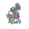
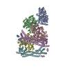
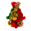
EMDB-19496: 
Structure of the formin Cdc12 bound to the barbed end of phalloidin-stabilized F-actin.
EMDB-19497:
Cryo-EM reconstruction of the formin Cdc12 bound to the barbed end of F-actin (without phalloidin)

EMDB-19499:
Structure of the F-actin barbed end bound by Cdc12 and profilin (ring complex) at a resolution of 6.3 Angstrom

EMDB-19501:
Structure of the undecorated barbed end of F-actin.

EMDB-19503:
Structure of the F-actin barbed end bound by formin mDia1

EMDB-19522:
Structure of the formin INF2 bound to the barbed end of F-actin.
PDB-8rtt:
Structure of the formin Cdc12 bound to the barbed end of phalloidin-stabilized F-actin.

PDB-8rty:
Structure of the F-actin barbed end bound by Cdc12 and profilin (ring complex) at a resolution of 6.3 Angstrom
PDB-8ru0:
Structure of the undecorated barbed end of F-actin.
PDB-8ru2:
Structure of the F-actin barbed end bound by formin mDia1
PDB-8rv2:
Structure of the formin INF2 bound to the barbed end of F-actin.
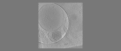
EMDB-18110:
The fibrillar and amorphous states of polyQ Q97
EMDB-18114:
phagophore in fibrillar polyQ
EMDB-18115:
phagophore and lysosomes with amorphous polyQ

EMDB-18116:
autolysosome, lysosome next to polyQ fibrils are empty
EMDB-18117:
autophagosome and autolysosomes are empty next to fibrillar polyQ
EMDB-18118:
Isolated autophagosome

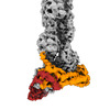
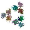
EMDB-16791:
Photorhabdus luminescens TcdA1 prepore-to-pore intermediate, K1179W mutant

EMDB-16792:
Photorhabdus luminescens TcdA1 prepore-to-pore intermediate, K567W K2008W mutant
EMDB-16793:
Photorhabdus luminescens TcdA1 prepore-to-pore intermediate, C16S, C20S, C870S, T1279C mutant
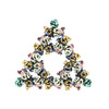
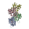
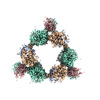
EMDB-19250:
Pseudoatomic model of a second-order Sierpinski triangle formed by the citrate synthase from Synechococcus elongatus
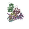
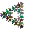
EMDB-19251:
Structure of a first order Sierpinski triangle formed by the H369R mutant of the citrate synthase from Synechococcus elongatus
PDB-8rjk:
Pseudoatomic model of a second-order Sierpinski triangle formed by the citrate synthase from Synechococcus elongatus
PDB-8rjl:
Structure of a first order Sierpinski triangle formed by the H369R mutant of the citrate synthase from Synechococcus elongatus
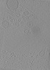
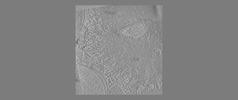
EMDB-35809:
Cellular components in INS-1E cell periphery
EMDB-35839:
Cellular components at INS-1E cell periphery under second phase of glucose-stimulated insulin secretion
EMDB-35840:
Cellular components at INS-1E cell periphery under first phase of glucose-stimulated insulin secretion
EMDB-35841:
Cellular components at INS-1E cell periphery under basal condition
EMDB-35842:
Cellular components at INS-1E cell periphery under basal condition

EMDB-35843:
Cellular components at INS-1E cell periphery under basal condition

EMDB-35844:
Cellular components at INS-1E cell periphery under basal condition
EMDB-35845:
Cellular components at INS-1E cell periphery under basal condition
EMDB-35846:
Cellular components at INS-1E cell periphery under basal condition
EMDB-35847:
Cellular components at INS-1E cell periphery under basal condition
EMDB-35848:
Cellular components at INS-1E cell periphery under second phase of glucose-stimulated insulin secretion
EMDB-35849:
Cellular components at INS-1E cell periphery under second phase of glucose-stimulated insulin secretion
EMDB-35850:
Cellular components at INS-1E cell periphery under second phase of glucose-stimulated insulin secretion
EMDB-35851:
Cellular components at INS-1E cell periphery under second phase of glucose-stimulated insulin secretion

EMDB-35852:
Cellular components at INS-1E cell periphery under second phase of glucose-stimulated insulin secretion
EMDB-35853:
Cellular components at INS-1E cell periphery under second phase of glucose-stimulated insulin secretion
EMDB-35854:
Cellular components at INS-1E cell periphery under second phase of glucose-stimulated insulin secretion
EMDB-35855:
Cellular components at INS-1E cell periphery under first phase of glucose-stimulated insulin secretion
EMDB-35856:
Cellular components at INS-1E cell periphery under first phase of glucose-stimulated insulin secretion
EMDB-35857:
Cellular components at INS-1E cell periphery under first phase of glucose-stimulated insulin secretion
EMDB-35858:
Cellular components at INS-1E cell periphery under first phase of glucose-stimulated insulin secretion
EMDB-35859:
Cellular components at INS-1E cell periphery under first phase of glucose-stimulated insulin secretion
EMDB-35860:
Cellular components at INS-1E cell periphery under first phase of glucose-stimulated insulin secretion
EMDB-35861:
Cellular components at INS-1E cell periphery under first phase of glucose-stimulated insulin secretion

EMDB-35874:
Cellular components at INS-1E cell interior under basal condition
EMDB-35875:
Cellular components at INS-1E cell interior under basal condition
Pages:
 wwPDB to switch to version 3 of the EMDB data model
