Journal: Science / Year: 2024 Title: Molecular mechanism of actin filament elongation by formins. Authors: Wout Oosterheert / Micaela Boiero Sanders / Johanna Funk / Daniel Prumbaum / Stefan Raunser / Peter Bieling / Abstract: Formins control the assembly of actin filaments (F-actin) that drive cell morphogenesis and motility in eukaryotes. However, their molecular interaction with F-actin and their mechanism of action ...Formins control the assembly of actin filaments (F-actin) that drive cell morphogenesis and motility in eukaryotes. However, their molecular interaction with F-actin and their mechanism of action remain unclear. In this work, we present high-resolution cryo-electron microscopy structures of F-actin barbed ends bound by three distinct formins, revealing a common asymmetric formin conformation imposed by the filament. Formation of new intersubunit contacts during actin polymerization sterically displaces formin and triggers its translocation. This "undock-and-lock" mechanism explains how actin-filament growth is coordinated with formin movement. Filament elongation speeds are controlled by the positioning and stability of actin-formin interfaces, which distinguish fast and slow formins. Furthermore, we provide a structure of the actin-formin-profilin ring complex, which resolves how profilin is rapidly released from the barbed end during filament elongation.
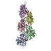
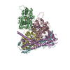
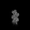
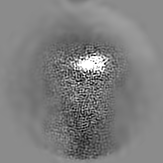
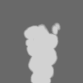
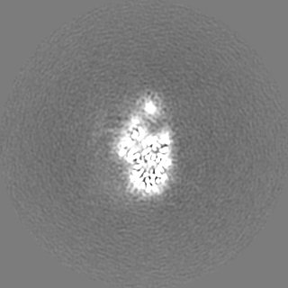
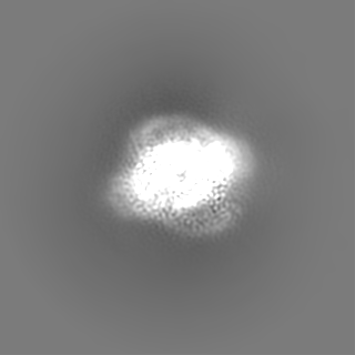
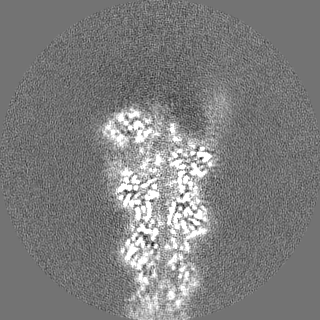
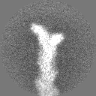
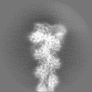
Unsharpened cryo-EM density map of actin-Cdc12. This reconstruction was computed with more particles, but displays weaker density for the FH2L domain of Cdc12 due to flexibility.
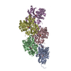
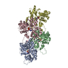
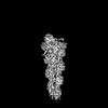
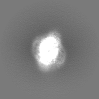
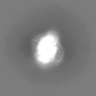
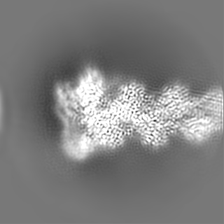
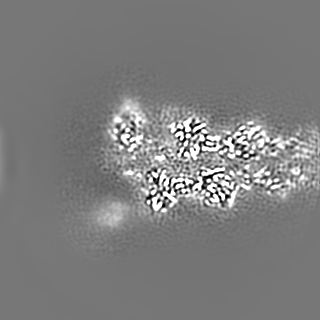
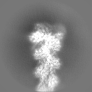
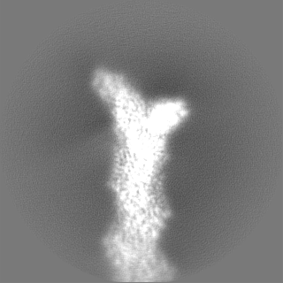
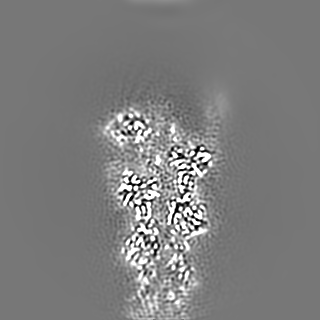
Composite map of two reconstructions of the formin Cdc12 bound to the barbed end of phalloidin-stabilized F-actin. This map was created using phenix and was used for visualization purposes.
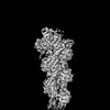
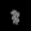
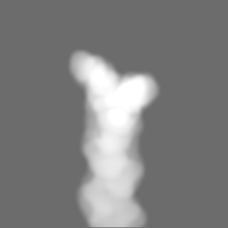
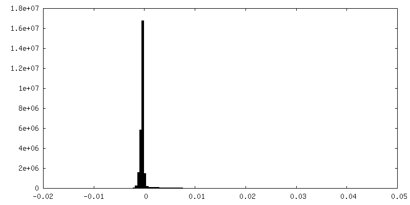
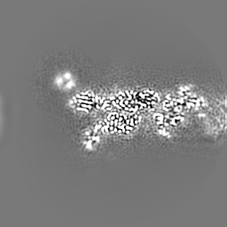
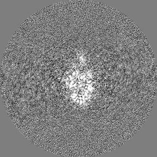
Unfiltered half map 1 of actin-Cdc12. This reconstruction was computed with more particles, but displays weaker density for the FH2L domain of Cdc12 due to flexibility.
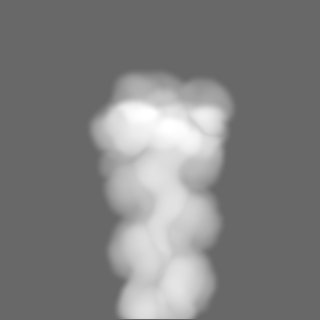
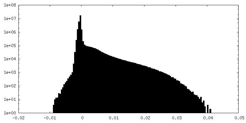
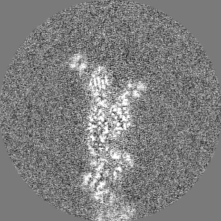
Unfiltered half map 2 of actin-Cdc12. This reconstruction was computed with more particles, but displays weaker density for the FH2L domain of Cdc12 due to flexibility.
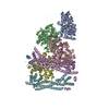
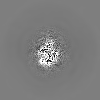
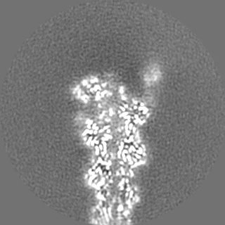
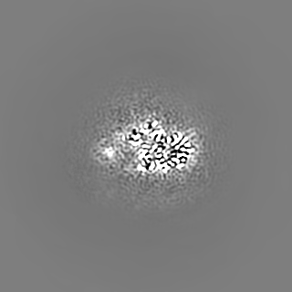
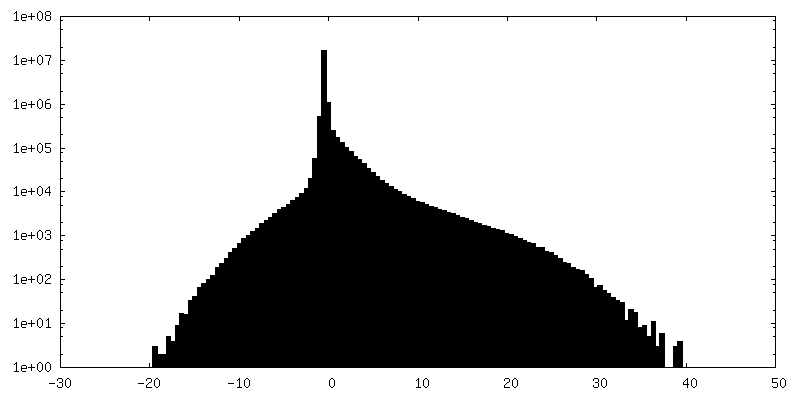
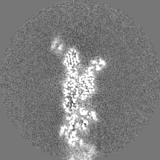
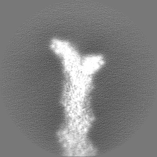
Sharpened density map of actin-Cdc12. This reconstruction was computed with more particles, but displays weaker density for the FH2L domain of Cdc12 due to flexibility.
Entire : Complex of the dimeric FH2 domain of S. Pombe Cdc12 bound to the ...
Entire
Name: Complex of the dimeric FH2 domain of S. Pombe Cdc12 bound to the barbed end of phalloidin stabilized F-actin.
Components
Complex: Complex of the dimeric FH2 domain of S. Pombe Cdc12 bound to the barbed end of phalloidin stabilized F-actin.
Complex: Actin filament
Protein or peptide: Actin, cytoplasmic 1, N-terminally processed
Complex: Dimeric FH2 domain of S. Pombe Cdc12
Protein or peptide: Cell division control protein 12
Complex: Phalloidin.
Protein or peptide: Phalloidin
Ligand: ADENOSINE-5'-DIPHOSPHATE
Ligand: MAGNESIUM ION
Ligand: PHOSPHATE ION
+
Supramolecule #1: Complex of the dimeric FH2 domain of S. Pombe Cdc12 bound to the ...
Supramolecule
Name: Complex of the dimeric FH2 domain of S. Pombe Cdc12 bound to the barbed end of phalloidin stabilized F-actin. type: complex / ID: 1 / Parent: 0 / Macromolecule list: #1-#3 Details: Human beta-actin and S. Pombe Cdc12 were purified separately, phalloidin (from Amanita phalloides) was bought from sigma. All components were mixed to assemble the complex prior to cryo-EM grid preparation.
Macromolecule #2: Cell division control protein 12
Macromolecule
Name: Cell division control protein 12 / type: protein_or_peptide / ID: 2 Details: FH2 domain of S. pombe Cdc12, purified from E. coli cells. Number of copies: 2 / Enantiomer: LEVO
In the structure databanks used in Yorodumi, some data are registered as the other names, "COVID-19 virus" and "2019-nCoV". Here are the details of the virus and the list of structure data.
Jan 31, 2019. EMDB accession codes are about to change! (news from PDBe EMDB page)
EMDB accession codes are about to change! (news from PDBe EMDB page)
The allocation of 4 digits for EMDB accession codes will soon come to an end. Whilst these codes will remain in use, new EMDB accession codes will include an additional digit and will expand incrementally as the available range of codes is exhausted. The current 4-digit format prefixed with “EMD-” (i.e. EMD-XXXX) will advance to a 5-digit format (i.e. EMD-XXXXX), and so on. It is currently estimated that the 4-digit codes will be depleted around Spring 2019, at which point the 5-digit format will come into force.
The EM Navigator/Yorodumi systems omit the EMD- prefix.
Related info.:Q: What is EMD? / ID/Accession-code notation in Yorodumi/EM Navigator
Yorodumi is a browser for structure data from EMDB, PDB, SASBDB, etc.
This page is also the successor to EM Navigator detail page, and also detail information page/front-end page for Omokage search.
The word "yorodu" (or yorozu) is an old Japanese word meaning "ten thousand". "mi" (miru) is to see.
Related info.:EMDB / PDB / SASBDB / Comparison of 3 databanks / Yorodumi Search / Aug 31, 2016. New EM Navigator & Yorodumi / Yorodumi Papers / Jmol/JSmol / Function and homology information / Changes in new EM Navigator and Yorodumi
 Movie
Movie Controller

 Yorodumi
 Open data
 Basic information
 Map data
 Sample
 Keywords
 Function and homology information
 Homo sapiens (human) /
 Amanita phalloides (death cap)
 Authors
 Germany, European Union, 3 items
  Citation
 Structure visualization
 Downloads & links
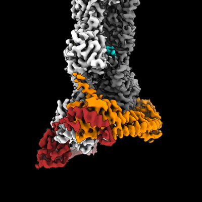 emd_19496.png
 http://ftp.pdbj.org/pub/emdb/structures/EMD-19496
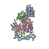


 Z (Sec.)
 Y (Row.)
 X (Col.)


 Sample components
 Trichoplusia ni (cabbage looper)


 Processing
 Electron microscopy
 FIELD EMISSION GUN