[English] 日本語
Yorodumi
- PDB-8a2s: Cryo-EM structure of F-actin in the Mg2+-ADP-Pi nucleotide state. -

+
Open data


ID or keywords:

Loading...

-
Basic information

Entry
Database: PDB / ID: 8a2s
TitleCryo-EM structure of F-actin in the Mg2+-ADP-Pi nucleotide state.
ComponentsActin, alpha skeletal muscle
KeywordsSTRUCTURAL PROTEIN / actin / cytoskeleton / filament / nucleotide state
Function / homology
Function and homology information


cytoskeletal motor activator activity / tropomyosin binding / myosin heavy chain binding / mesenchyme migration / troponin I binding / actin filament bundle / filamentous actin / actin filament bundle assembly / skeletal muscle thin filament assembly / striated muscle thin filament ...cytoskeletal motor activator activity / tropomyosin binding / myosin heavy chain binding / mesenchyme migration / troponin I binding / actin filament bundle / filamentous actin / actin filament bundle assembly / skeletal muscle thin filament assembly / striated muscle thin filament / skeletal muscle myofibril / actin monomer binding / skeletal muscle fiber development / stress fiber / titin binding / actin filament polymerization / filopodium / actin filament / Hydrolases; Acting on acid anhydrides; Acting on acid anhydrides to facilitate cellular and subcellular movement / calcium-dependent protein binding / lamellipodium / cell body / hydrolase activity / protein domain specific binding / calcium ion binding / positive regulation of gene expression / magnesium ion binding / ATP binding / identical protein binding / cytoplasm
Similarity search - Function
Actins signature 1. / Actin, conserved site / Actins signature 2. / Actin/actin-like conserved site / Actins and actin-related proteins signature. / Actin / Actin family / Actin / ATPase, nucleotide binding domain
Similarity search - Domain/homology
ADENOSINE-5'-DIPHOSPHATE / PHOSPHATE ION / Actin, alpha skeletal muscle
Similarity search - Component
Biological speciesOryctolagus cuniculus (rabbit)
MethodELECTRON MICROSCOPY / single particle reconstruction / cryo EM / Resolution: 2.22 Å
AuthorsOosterheert, W. / Klink, B.U. / Belyy, A. / Pospich, S. / Raunser, S.
Funding supportEuropean Union, Germany, 4items
OrganizationGrant numberCountry
European Research Council (ERC)856118European Union
Alexander von Humboldt Foundation Germany
European Molecular Biology Organization (EMBO)European Union
Max Planck Society Germany
Citation
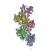
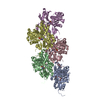
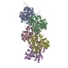
Journal: Nature / Year: 2022
Title: Structural basis of actin filament assembly and aging.
Authors: Wout Oosterheert / Björn U Klink / Alexander Belyy / Sabrina Pospich / Stefan Raunser /
Abstract: The dynamic turnover of actin filaments (F-actin) controls cellular motility in eukaryotes and is coupled to changes in the F-actin nucleotide state. It remains unclear how F-actin hydrolyses ATP and ...The dynamic turnover of actin filaments (F-actin) controls cellular motility in eukaryotes and is coupled to changes in the F-actin nucleotide state. It remains unclear how F-actin hydrolyses ATP and subsequently undergoes subtle conformational rearrangements that ultimately lead to filament depolymerization by actin-binding proteins. Here we present cryo-electron microscopy structures of F-actin in all nucleotide states, polymerized in the presence of Mg or Ca at approximately 2.2 Å resolution. The structures show that actin polymerization induces the relocation of water molecules in the nucleotide-binding pocket, activating one of them for the nucleophilic attack of ATP. Unexpectedly, the back door for the subsequent release of inorganic phosphate (P) is closed in all structures, indicating that P release occurs transiently. The small changes in the nucleotide-binding pocket after ATP hydrolysis and P release are sensed by a key amino acid, amplified and transmitted to the filament periphery. Furthermore, differences in the positions of water molecules in the nucleotide-binding pocket explain why Ca-actin shows slower polymerization rates than Mg-actin. Our work elucidates the solvent-driven rearrangements that govern actin filament assembly and aging and lays the foundation for the rational design of drugs and small molecules for imaging and therapeutic applications.
History
DepositionJun 6, 2022Deposition site: PDBE / Processing site: PDBE
Revision 1.0Aug 10, 2022Provider: repository / Type: Initial release
Revision 1.1Nov 16, 2022Group: Database references / Category: citation / citation_author
Item: _citation.country / _citation.journal_abbrev ..._citation.country / _citation.journal_abbrev / _citation.journal_id_ASTM / _citation.journal_id_CSD / _citation.journal_id_ISSN / _citation.pdbx_database_id_DOI / _citation.pdbx_database_id_PubMed / _citation.title / _citation.year / _citation_author.identifier_ORCID
Revision 1.2Nov 23, 2022Group: Database references / Category: citation / citation_author
Item: _citation.journal_volume / _citation.page_first ..._citation.journal_volume / _citation.page_first / _citation.page_last / _citation_author.identifier_ORCID

-
Structure visualization

Structure viewerMolecule:
MolmilJmol/JSmol

Downloads & links

-
Assembly

Deposited unit
C: Actin, alpha skeletal muscle
A: Actin, alpha skeletal muscle
B: Actin, alpha skeletal muscle
D: Actin, alpha skeletal muscle
E: Actin, alpha skeletal muscle
hetero molecules


Theoretical massNumber of molelcules
Total (without water)212,11120
Polymers209,3785
Non-polymers2,73215
Water10,647591
1


  • Idetical with deposited unit
  • defined by author
  • Evidence: electron microscopy
TypeNameSymmetry operationNumber
identity operation1_5551
Noncrystallographic symmetry (NCS)NCS domain:
IDEns-IDDetails
d_1ens_1chain "A"
d_2ens_1chain "B"
d_3ens_1chain "C"
d_4ens_1chain "D"
d_5ens_1chain "E"

NCS domain segments:
Dom-IDComponent-IDEns-IDBeg label comp-IDEnd label comp-IDLabel asym-IDLabel seq-ID
d_11ens_1THRPHEF1 - 367
d_12ens_1ADPADPG
d_13ens_1MGMGH
d_14ens_1PO4PO4I
d_21ens_1THRPHEK1 - 367
d_22ens_1ADPADPL
d_23ens_1MGMGM
d_24ens_1PO4PO4N
d_31ens_1THRPHEA1 - 367
d_32ens_1ADPADPB
d_33ens_1MGMGC
d_34ens_1PO4PO4D
d_41ens_1THRPHEP1 - 367
d_42ens_1ADPADPQ
d_43ens_1MGMGR
d_44ens_1PO4PO4S
d_51ens_1THRPHEU1 - 367
d_52ens_1ADPADPV
d_53ens_1MGMGW
d_54ens_1PO4PO4X

NCS oper:
IDCodeMatrixVector
1given(-0.957466187451, 0.222016384287, -0.184301994011), (-0.241868142939, -0.96583744649, 0.0930474523532), (-0.15734770834, 0.133666570488, 0.978455388157)252.586728275, 280.471026861, 33.4660852884
2given(0.891694111748, -0.452375538951, 0.0154266922433), (0.451327489312, 0.891186986345, 0.0457083445472), (-0.0344254043686, -0.0337953714119, 0.99883570441)67.0892579067, -48.5346305365, 63.9234615613
3given(-0.748425680092, 0.635598356507, -0.189403618194), (-0.653201160816, -0.7558714813, 0.0445706996304), (-0.114835730015, 0.15707651945, 0.980887211737)166.322855269, 316.942986731, 79.1349639926
4given(0.592603358682, -0.805429635522, 0.0102157477613), (0.801398598353, 0.590819986051, 0.0932310604929), (-0.081126727022, -0.0470621536445, 0.995592089089)149.174847866, -58.5963284745, 126.955968553

-
Components

#1: Protein
Actin, alpha skeletal muscle / / Alpha-actin-1


Mass: 41875.633 Da / Num. of mol.: 5 / Source method: isolated from a natural source / Source: (natural) Oryctolagus cuniculus (rabbit) / Plasmid details: purified from muscle acetone powder / Tissue: skeletal muscle / References: UniProt: P68135
#2: Chemical
ChemComp-ADP / ADENOSINE-5'-DIPHOSPHATE / Adenosine diphosphate


Mass: 427.201 Da / Num. of mol.: 5 / Source method: obtained synthetically / Formula: C10H15N5O10P2 / Feature type: SUBJECT OF INVESTIGATION / Comment: ADP, energy-carrying molecule*YM
#3: Chemical
ChemComp-MG / MAGNESIUM ION


Mass: 24.305 Da / Num. of mol.: 5 / Source method: obtained synthetically / Formula: Mg / Feature type: SUBJECT OF INVESTIGATION
#4: Chemical
ChemComp-PO4 / PHOSPHATE ION / Phosphate


Mass: 94.971 Da / Num. of mol.: 5 / Source method: obtained synthetically / Formula: PO4 / Feature type: SUBJECT OF INVESTIGATION
#5: Water ChemComp-HOH / water / Water


Mass: 18.015 Da / Num. of mol.: 591 / Source method: isolated from a natural source / Formula: H2O
Has ligand of interestY

-
Experimental details

-
Experiment

ExperimentMethod: ELECTRON MICROSCOPY
EM experimentAggregation state: FILAMENT / 3D reconstruction method: single particle reconstruction

-
Sample preparation

ComponentName: rabbit skeletal alpha-actin in the filamentous state. / Type: COMPLEX
Details: The helical rise of F-actin is 27.5 Angstrom, with a helical twist of ~166.5 degrees.
Entity ID: #1 / Source: NATURAL
Molecular weightValue: 15.2 kDa/nm / Experimental value: NO
Source (natural)Organism: Oryctolagus cuniculus (rabbit) / Organ: skeletal muscle
Buffer solutionpH: 7.5
Details: F-phosphate buffer: 5 mM Tris, 100 mM KCl, 2 mM MgCl2, 2 mM NaN3, 1 mM DTT, 50 mM potassium phosphate pH 7.5
Buffer component
IDConc.NameFormulaBuffer-ID
15 mMTris1
250 mMpotassium chlorideKCl1
32 mMmagnesium chlorideMgCl21
42 mMsodium azideNaN31
51 mMdithiothreitol1
650 mMpotassium phosphate1
SpecimenConc.: 0.67 mg/ml / Embedding applied: NO / Shadowing applied: NO / Staining applied: NO / Vitrification applied: YES
Specimen supportGrid material: COPPER / Grid mesh size: 300 divisions/in. / Grid type: Quantifoil R2/1
VitrificationInstrument: FEI VITROBOT MARK IV / Cryogen name: ETHANE-PROPANE / Humidity: 100 % / Chamber temperature: 286 K
Details: The Vitrobot was operated at 13 degrees celsius and the samples were blotted for 9 seconds with a blot force of -25.

-
Electron microscopy imaging

Experimental equipment
Model: Titan Krios / Image courtesy: FEI Company
MicroscopyModel: FEI TITAN KRIOS
Electron gunElectron source: FIELD EMISSION GUN / Accelerating voltage: 300 kV / Illumination mode: FLOOD BEAM
Electron lensMode: BRIGHT FIELDBright-field microscopy / Nominal magnification: 130000 X / Nominal defocus max: 2000 nm / Nominal defocus min: 700 nm / Cs: 2.7 mm / C2 aperture diameter: 50 µm / Alignment procedure: ZEMLIN TABLEAU
Specimen holderCryogen: NITROGEN / Specimen holder model: FEI TITAN KRIOS AUTOGRID HOLDER
Image recordingAverage exposure time: 3 sec. / Electron dose: 88.8 e/Å2 / Film or detector model: GATAN K3 BIOQUANTUM (6k x 4k) / Num. of grids imaged: 1 / Num. of real images: 9658 / Details: Images were collected in supperresolution mode.
EM imaging opticsEnergyfilter slit width: 15 eV
Image scansWidth: 5760 / Height: 4092

-
Processing

Software
NameVersionClassificationNB
phenix.real_space_refine1.20.1_4487refinement
PHENIX1.20.1_4487refinement
EM software
IDNameVersionCategoryDetails
1crYOLO1.8particle selection
2EPUimage acquisition4-5 images per hole.
4CTFFIND4.13CTF correction
7UCSF ChimeraX1.3model fitting
9PHENIXv1.20.1-4487model refinementreal space refinement
10SPHIRE1.4initial Euler assignment
11RELION3.1.0final Euler assignment
12RELION3.1.0classification
13RELION3.1.03D reconstruction
CTF correctionType: PHASE FLIPPING AND AMPLITUDE CORRECTION
Particle selectionNum. of particles selected: 2349979
Details: We picked helical segments with a box distance of 40 pixels or 27.8 Angstrom and a minimum number of six boxes per filament.
SymmetryPoint symmetry: C1 (asymmetric)
3D reconstructionResolution: 2.22 Å / Resolution method: FSC 0.143 CUT-OFF / Num. of particles: 1808554 / Symmetry type: POINT
Atomic model buildingProtocol: FLEXIBLE FIT / Space: REAL
Details: The structures were then refined through a similar protocol of iterative cycles in Coot and phenix real-space refine. All solvent molecules (ions, waters) were placed manually in Coot in the ...Details: The structures were then refined through a similar protocol of iterative cycles in Coot and phenix real-space refine. All solvent molecules (ions, waters) were placed manually in Coot in the central actin subunit, and were then placed in the other subunits using NCS. Because the local resolution of each F-actin reconstruction is highest in the center and lower at the periphery of the map, we inspected all waters in each structure manually before the final phenix refinement; water molecular with poor corresponding cryo-EM density were removed.
Atomic model buildingPDB-ID: 7AHN
RefinementCross valid method: NONE
Stereochemistry target values: GeoStd + Monomer Library + CDL v1.2
Displacement parametersBiso mean: 34.08 Å2
Refine LS restraints
Refine-IDTypeDev idealNumber
ELECTRON MICROSCOPYf_bond_d0.008314860
ELECTRON MICROSCOPYf_angle_d0.665820170
ELECTRON MICROSCOPYf_chiral_restr0.05312240
ELECTRON MICROSCOPYf_plane_restr0.00542570
ELECTRON MICROSCOPYf_dihedral_angle_d17.33115520
Refine LS restraints NCS
Ens-IDDom-IDAuth asym-IDRefine-IDTypeRms dev position (Å)
ens_1d_2AELECTRON MICROSCOPYNCS constraints7.02933071242E-13
ens_1d_3AELECTRON MICROSCOPYNCS constraints8.73804946678E-13
ens_1d_4AELECTRON MICROSCOPYNCS constraints1.80703718994E-11
ens_1d_5AELECTRON MICROSCOPYNCS constraints6.53802392143E-13

+
About Yorodumi

-
News

-
Feb 9, 2022. New format data for meta-information of EMDB entries

New format data for meta-information of EMDB entries

  • Version 3 of the EMDB header file is now the official format.
  • The previous official version 1.9 will be removed from the archive.

Related info.:EMDB header

External links:wwPDB to switch to version 3 of the EMDB data model

-
Aug 12, 2020. Covid-19 info

Covid-19 info

URL: https://pdbj.org/emnavi/covid19.php

New page: Covid-19 featured information page in EM Navigator.

Related info.:Covid-19 info / Mar 5, 2020. Novel coronavirus structure data

+
Mar 5, 2020. Novel coronavirus structure data

Novel coronavirus structure data

Related info.:Yorodumi Speices / Aug 12, 2020. Covid-19 info

External links:COVID-19 featured content - PDBj / Molecule of the Month (242):Coronavirus Proteases

+
Jan 31, 2019. EMDB accession codes are about to change! (news from PDBe EMDB page)

EMDB accession codes are about to change! (news from PDBe EMDB page)

  • The allocation of 4 digits for EMDB accession codes will soon come to an end. Whilst these codes will remain in use, new EMDB accession codes will include an additional digit and will expand incrementally as the available range of codes is exhausted. The current 4-digit format prefixed with “EMD-” (i.e. EMD-XXXX) will advance to a 5-digit format (i.e. EMD-XXXXX), and so on. It is currently estimated that the 4-digit codes will be depleted around Spring 2019, at which point the 5-digit format will come into force.
  • The EM Navigator/Yorodumi systems omit the EMD- prefix.

Related info.:Q: What is EMD? / ID/Accession-code notation in Yorodumi/EM Navigator

External links:EMDB Accession Codes are Changing Soon! / Contact to PDBj

+
Jul 12, 2017. Major update of PDB

Major update of PDB

  • wwPDB released updated PDB data conforming to the new PDBx/mmCIF dictionary.
  • This is a major update changing the version number from 4 to 5, and with Remediation, in which all the entries are updated.
  • In this update, many items about electron microscopy experimental information are reorganized (e.g. em_software).
  • Now, EM Navigator and Yorodumi are based on the updated data.

External links:wwPDB Remediation / Enriched Model Files Conforming to OneDep Data Standards Now Available in the PDB FTP Archive

-
Yorodumi

Thousand views of thousand structures

  • Yorodumi is a browser for structure data from EMDB, PDB, SASBDB, etc.
  • This page is also the successor to EM Navigator detail page, and also detail information page/front-end page for Omokage search.
  • The word "yorodu" (or yorozu) is an old Japanese word meaning "ten thousand". "mi" (miru) is to see.

Related info.:EMDB / PDB / SASBDB / Comparison of 3 databanks / Yorodumi Search / Aug 31, 2016. New EM Navigator & Yorodumi / Yorodumi Papers / Jmol/JSmol / Function and homology information / Changes in new EM Navigator and Yorodumi

Read more