 Movie
Movie Controller

[English] 日本語
 Yorodumi
- PDB-8rjk: Pseudoatomic model of a second-order Sierpinski triangle formed b... -
+ Open data

- Basic information
| Entry | Database: PDB / ID: 8rjk | ||||||||||||
|---|---|---|---|---|---|---|---|---|---|---|---|---|---|
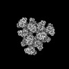
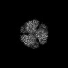
| Title | Pseudoatomic model of a second-order Sierpinski triangle formed by the citrate synthase from Synechococcus elongatus | ||||||||||||
 Components Components | Citrate synthase | ||||||||||||
 Keywords Keywords | TRANSFERASE / Fractal complex | ||||||||||||
| Function / homology |  Function and homology information Function and homology informationcitrate synthase activity / tricarboxylic acid cycle / carbohydrate metabolic process / metal ion binding / cytosol Similarity search - Function | ||||||||||||
| Biological species |  Synechococcus elongatus PCC 7942 = FACHB-805 (bacteria) Synechococcus elongatus PCC 7942 = FACHB-805 (bacteria) | ||||||||||||
| Method | ELECTRON MICROSCOPY / single particle reconstruction / cryo EM / Resolution: 5.91 Å | ||||||||||||
 Authors Authors | Lo, Y.K. / Bohn, S. / Sendker, F.L. / Schuller, J.M. / Hochberg, G. | ||||||||||||
| Funding support |  Germany, European Union, 3items Germany, European Union, 3items
| ||||||||||||
 Citation Citation | Journal: Nature / Year: 2024 Title: Emergence of fractal geometries in the evolution of a metabolic enzyme. Authors: Franziska L Sendker / Yat Kei Lo / Thomas Heimerl / Stefan Bohn / Louise J Persson / Christopher-Nils Mais / Wiktoria Sadowska / Nicole Paczia / Eva Nußbaum / María Del Carmen Sánchez ...Authors: Franziska L Sendker / Yat Kei Lo / Thomas Heimerl / Stefan Bohn / Louise J Persson / Christopher-Nils Mais / Wiktoria Sadowska / Nicole Paczia / Eva Nußbaum / María Del Carmen Sánchez Olmos / Karl Forchhammer / Daniel Schindler / Tobias J Erb / Justin L P Benesch / Erik G Marklund / Gert Bange / Jan M Schuller / Georg K A Hochberg /   Abstract: Fractals are patterns that are self-similar across multiple length-scales. Macroscopic fractals are common in nature; however, so far, molecular assembly into fractals is restricted to synthetic ...Fractals are patterns that are self-similar across multiple length-scales. Macroscopic fractals are common in nature; however, so far, molecular assembly into fractals is restricted to synthetic systems. Here we report the discovery of a natural protein, citrate synthase from the cyanobacterium Synechococcus elongatus, which self-assembles into Sierpiński triangles. Using cryo-electron microscopy, we reveal how the fractal assembles from a hexameric building block. Although different stimuli modulate the formation of fractal complexes and these complexes can regulate the enzymatic activity of citrate synthase in vitro, the fractal may not serve a physiological function in vivo. We use ancestral sequence reconstruction to retrace how the citrate synthase fractal evolved from non-fractal precursors, and the results suggest it may have emerged as a harmless evolutionary accident. Our findings expand the space of possible protein complexes and demonstrate that intricate and regulatable assemblies can evolve in a single substitution. | ||||||||||||
| History |
|
- Structure visualization
| Structure viewer | Molecule: MolmilJmol/JSmol |
|---|
- Downloads & links
-Download
| PDBx/mmCIF format | 8rjk.cif.gz | 2.5 MB | Display | PDBx/mmCIF format |
|---|---|---|---|---|
| PDB format | pdb8rjk.ent.gz | 1.8 MB | Display | PDB format |
| PDBx/mmJSON format | 8rjk.json.gz | Tree view | PDBx/mmJSON format | |
| Others |  Other downloads Other downloads |
-Validation report
| Arichive directory | https://data.pdbj.org/pub/pdb/validation_reports/rj/8rjkftp://data.pdbj.org/pub/pdb/validation_reports/rj/8rjk | HTTPS FTP |
|---|
-Related structure data
| Related structure data |  19250MC 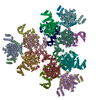 8an1C 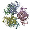 8beiC  8bp7C 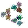 8rjlC M: map data used to model this data C: citing same article ( |
|---|---|
| Similar structure data |

-Links
 PDBj
PDBj

- Assembly
| Deposited unit | 
|
|---|---|
| 1 |
|
-Components
| #1: Protein | Mass: 44386.422 Da / Num. of mol.: 54 / Mutation: H369R Source method: isolated from a genetically manipulated source Source: (gene. exp.) Synechococcus elongatus PCC 7942 = FACHB-805 (bacteria)Gene: Synpcc7942_0612 / Production host: |
|---|
-Experimental details
-Experiment
| Experiment | Method: ELECTRON MICROSCOPY |
|---|---|
| EM experiment | Aggregation state: PARTICLE / 3D reconstruction method: single particle reconstruction |
- Sample preparation
| Component | Name: Second order Sierpinski triangle formed by the citrate synthase from Synechoccocus elongatus Type: COMPLEX / Entity ID: all / Source: RECOMBINANT |
|---|---|
| Source (natural) | Organism: Synechococcus elongatus PCC 7942 = FACHB-805 (bacteria) |
| Source (recombinant) | Organism: |
| Buffer solution | pH: 7.5 |
| Specimen | Embedding applied: NO / Shadowing applied: NO / Staining applied: NO / Vitrification applied: YES |
| Vitrification | Cryogen name: ETHANE-PROPANE / Humidity: 100 % / Chamber temperature: 277 K |
- Electron microscopy imaging
| Experimental equipment |  Model: Titan Krios / Image courtesy: FEI Company |
|---|---|
| Microscopy | Model: FEI TITAN KRIOS |
| Electron gun | Electron source:  FIELD EMISSION GUN / Accelerating voltage: 300 kV / Illumination mode: FLOOD BEAM FIELD EMISSION GUN / Accelerating voltage: 300 kV / Illumination mode: FLOOD BEAM |
| Electron lens | Mode: BRIGHT FIELD / Nominal defocus max: 3000 nm / Nominal defocus min: 500 nm |
| Image recording | Electron dose: 60 e/Å2 / Film or detector model: FEI FALCON IV (4k x 4k) |
- Processing
| CTF correction | Type: PHASE FLIPPING AND AMPLITUDE CORRECTION |
|---|---|
| 3D reconstruction | Resolution: 5.91 Å / Resolution method: FSC 0.143 CUT-OFF / Num. of particles: 17191 / Symmetry type: POINT |
