 Movie
Movie Controller

[English] 日本語
 Yorodumi
- EMDB-19250: Pseudoatomic model of a second-order Sierpinski triangle formed b... -
+ Open data

- Basic information
| Entry |  | ||||||||||||
|---|---|---|---|---|---|---|---|---|---|---|---|---|---|
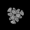
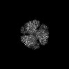
| Title | Pseudoatomic model of a second-order Sierpinski triangle formed by the citrate synthase from Synechococcus elongatus | ||||||||||||
 Map data Map data | |||||||||||||
 Sample Sample |
| ||||||||||||
 Keywords Keywords | Fractal complex / TRANSFERASE | ||||||||||||
| Function / homology |  Function and homology information Function and homology informationcitrate synthase activity / tricarboxylic acid cycle / carbohydrate metabolic process / metal ion binding / cytosol Similarity search - Function | ||||||||||||
| Biological species |  Synechococcus elongatus PCC 7942 = FACHB-805 (bacteria) Synechococcus elongatus PCC 7942 = FACHB-805 (bacteria) | ||||||||||||
| Method | single particle reconstruction / cryo EM / Resolution: 5.91 Å | ||||||||||||
 Authors Authors | Lo YK / Bohn S / Sendker FL / Schuller JM / Hochberg G | ||||||||||||
| Funding support |  Germany, European Union, 3 items Germany, European Union, 3 items
| ||||||||||||
 Citation Citation | Journal: Nature / Year: 2024 Title: Emergence of fractal geometries in the evolution of a metabolic enzyme. Authors: Franziska L Sendker / Yat Kei Lo / Thomas Heimerl / Stefan Bohn / Louise J Persson / Christopher-Nils Mais / Wiktoria Sadowska / Nicole Paczia / Eva Nußbaum / María Del Carmen Sánchez ...Authors: Franziska L Sendker / Yat Kei Lo / Thomas Heimerl / Stefan Bohn / Louise J Persson / Christopher-Nils Mais / Wiktoria Sadowska / Nicole Paczia / Eva Nußbaum / María Del Carmen Sánchez Olmos / Karl Forchhammer / Daniel Schindler / Tobias J Erb / Justin L P Benesch / Erik G Marklund / Gert Bange / Jan M Schuller / Georg K A Hochberg /   Abstract: Fractals are patterns that are self-similar across multiple length-scales. Macroscopic fractals are common in nature; however, so far, molecular assembly into fractals is restricted to synthetic ...Fractals are patterns that are self-similar across multiple length-scales. Macroscopic fractals are common in nature; however, so far, molecular assembly into fractals is restricted to synthetic systems. Here we report the discovery of a natural protein, citrate synthase from the cyanobacterium Synechococcus elongatus, which self-assembles into Sierpiński triangles. Using cryo-electron microscopy, we reveal how the fractal assembles from a hexameric building block. Although different stimuli modulate the formation of fractal complexes and these complexes can regulate the enzymatic activity of citrate synthase in vitro, the fractal may not serve a physiological function in vivo. We use ancestral sequence reconstruction to retrace how the citrate synthase fractal evolved from non-fractal precursors, and the results suggest it may have emerged as a harmless evolutionary accident. Our findings expand the space of possible protein complexes and demonstrate that intricate and regulatable assemblies can evolve in a single substitution. | ||||||||||||
| History |
|
- Structure visualization
| Supplemental images |
|---|

- Downloads & links
-EMDB archive
| Map data | emd_19250.map.gz | 778.6 MB | EMDB map data format | |
|---|---|---|---|---|
| Header (meta data) | emd-19250-v30.xmlemd-19250.xml | 14.7 KB 14.7 KB | Display Display | EMDB header |
| Images | 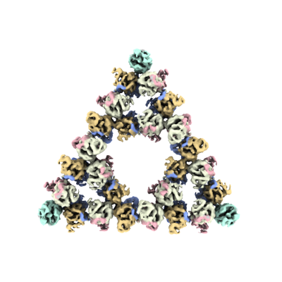 emd_19250.png emd_19250.png | 91.8 KB | ||
| Filedesc metadata | emd-19250.cif.gz | 5.6 KB | ||
| Others | emd_19250_half_map_1.map.gzemd_19250_half_map_2.map.gz | 758 MB 757.8 MB | ||
| Archive directory |  http://ftp.pdbj.org/pub/emdb/structures/EMD-19250ftp://ftp.pdbj.org/pub/emdb/structures/EMD-19250 http://ftp.pdbj.org/pub/emdb/structures/EMD-19250ftp://ftp.pdbj.org/pub/emdb/structures/EMD-19250 | HTTPS FTP |
-Related structure data
| Related structure data |  8rjkMC  8an1C  8beiC  8bp7C 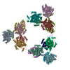 8rjlC M: atomic model generated by this map C: citing same article ( |
|---|---|
| Similar structure data |

-Links
| EMDB pages | EMDB (EBI/PDBe) / EMDataResource |
|---|---|
| Related items in Molecule of the Month |


-Map
| File | Download / File: emd_19250.map.gz / Format: CCP4 / Size: 824 MB / Type: IMAGE STORED AS FLOATING POINT NUMBER (4 BYTES) | ||||||||||||||||||||||||||||||||||||
|---|---|---|---|---|---|---|---|---|---|---|---|---|---|---|---|---|---|---|---|---|---|---|---|---|---|---|---|---|---|---|---|---|---|---|---|---|---|
| Projections & slices | Image control
Images are generated by Spider. | ||||||||||||||||||||||||||||||||||||
| Voxel size | X=Y=Z: 1.58 Å | ||||||||||||||||||||||||||||||||||||
| Density |
| ||||||||||||||||||||||||||||||||||||
| Symmetry | Space group: 1 | ||||||||||||||||||||||||||||||||||||
| Details | EMDB XML:
|
 Z (Sec.)
 Y (Row.)
 X (Col.)


-Supplemental data
-Half map: #2
| File | emd_19250_half_map_1.map | ||||||||||||
|---|---|---|---|---|---|---|---|---|---|---|---|---|---|
| Projections & Slices |
| ||||||||||||
| Density Histograms |


-Half map: #1
| File | emd_19250_half_map_2.map | ||||||||||||
|---|---|---|---|---|---|---|---|---|---|---|---|---|---|
| Projections & Slices |
| ||||||||||||
| Density Histograms |


- Sample components
-Entire : Second order Sierpinski triangle formed by the citrate synthase f...
| Entire | Name: Second order Sierpinski triangle formed by the citrate synthase from Synechoccocus elongatus |
|---|---|
| Components |
|
-Supramolecule #1: Second order Sierpinski triangle formed by the citrate synthase f...
| Supramolecule | Name: Second order Sierpinski triangle formed by the citrate synthase from Synechoccocus elongatus type: complex / ID: 1 / Parent: 0 / Macromolecule list: all |
|---|---|
| Source (natural) | Organism: Synechococcus elongatus PCC 7942 = FACHB-805 (bacteria) |
-Macromolecule #1: Citrate synthase
| Macromolecule | Name: Citrate synthase / type: protein_or_peptide / ID: 1 / Number of copies: 54 / Enantiomer: LEVO |
|---|---|
| Source (natural) | Organism: Synechococcus elongatus PCC 7942 = FACHB-805 (bacteria) |
| Molecular weight | Theoretical: 44.386422 KDa |
| Recombinant expression | Organism: |
| Sequence | String: MTAVSEFRPG LEGVPATLSS ISFVDGQRGV LEYRGISIEQ LAQQSSFLET AYLLIWGHLP TQQELTEFEH EIRYHRRIKF RIRDMMKCF PDSGHPMDAL QASAAALGLF YSRRALDDPE YIRAAVVRLL AKIPTMVAAF QLIRKGNDPI QPRDELDYAA N FLYMLTER ...String: MTAVSEFRPG LEGVPATLSS ISFVDGQRGV LEYRGISIEQ LAQQSSFLET AYLLIWGHLP TQQELTEFEH EIRYHRRIKF RIRDMMKCF PDSGHPMDAL QASAAALGLF YSRRALDDPE YIRAAVVRLL AKIPTMVAAF QLIRKGNDPI QPRDELDYAA N FLYMLTER EPDPVAARIF DICLTLHAEH TINASTFSAM VTASTLTDPY AVVASAVGTL AGPLHGGANE EVLDMLEAIG SV ENVEPYL DHCIATKTRI MGFGHRVYKV KDPRAVILQN LAEQLFDIFG HDPYYEIAVA VEKAAAERLS HKGIYPNVDF YSG LVYRKL GIPSDLFTPV FAIARVAGWL AHWKEQLNEN RIFRPTQIYT GSRNLDYTPI ADRDLAIESD LEHHHHHH UniProtKB: Citrate synthase |
-Experimental details
-Structure determination
| Method | cryo EM |
|---|---|
 Processing Processing | single particle reconstruction |
| Aggregation state | particle |
-Sample preparation
| Buffer | pH: 7.5 |
|---|---|
| Vitrification | Cryogen name: ETHANE-PROPANE / Chamber humidity: 100 % / Chamber temperature: 277 K |
- Electron microscopy
| Microscope | FEI TITAN KRIOS |
|---|---|
| Image recording | Film or detector model: FEI FALCON IV (4k x 4k) / Average electron dose: 60.0 e/Å2 |
| Electron beam | Acceleration voltage: 300 kV / Electron source:  FIELD EMISSION GUN FIELD EMISSION GUN |
| Electron optics | Illumination mode: FLOOD BEAM / Imaging mode: BRIGHT FIELD / Nominal defocus max: 3.0 µm / Nominal defocus min: 0.5 µm |
| Experimental equipment |  Model: Titan Krios / Image courtesy: FEI Company |
-Image processing
| Startup model | Type of model: NONE |
|---|---|
| Final reconstruction | Resolution.type: BY AUTHOR / Resolution: 5.91 Å / Resolution method: FSC 0.143 CUT-OFF / Number images used: 17191 |
| Initial angle assignment | Type: ANGULAR RECONSTITUTION |
| Final angle assignment | Type: ANGULAR RECONSTITUTION |
