Movie
Movie Controller

[English] 日本語
 Yorodumi
- PDB-3jd7: The novel asymmetric entry intermediate of a picornavirus capture... -
+ Open data

- Basic information
| Entry | Database: PDB / ID: 3jd7 | ||||||
|---|---|---|---|---|---|---|---|
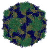
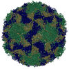
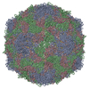
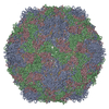
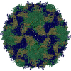
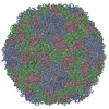
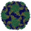
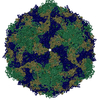
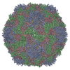
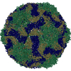
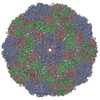
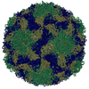
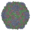
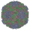
| Title | The novel asymmetric entry intermediate of a picornavirus captured with nanodiscs | ||||||
 Components Components |
| ||||||
 Keywords Keywords | VIRUS / picornavirus / entry intermediate | ||||||
| Function / homology |  Function and homology information Function and homology informationRNA-protein covalent cross-linking / : / : / symbiont-mediated suppression of host cytoplasmic pattern recognition receptor signaling pathway via inhibition of RIG-I activity / symbiont-mediated suppression of host cytoplasmic pattern recognition receptor signaling pathway via inhibition of MDA-5 activity / symbiont-mediated suppression of host cytoplasmic pattern recognition receptor signaling pathway via inhibition of MAVS activity / picornain 2A / symbiont-mediated suppression of host mRNA export from nucleus / symbiont genome entry into host cell via pore formation in plasma membrane / picornain 3C ...RNA-protein covalent cross-linking / : / : / symbiont-mediated suppression of host cytoplasmic pattern recognition receptor signaling pathway via inhibition of RIG-I activity / symbiont-mediated suppression of host cytoplasmic pattern recognition receptor signaling pathway via inhibition of MDA-5 activity / symbiont-mediated suppression of host cytoplasmic pattern recognition receptor signaling pathway via inhibition of MAVS activity / picornain 2A / symbiont-mediated suppression of host mRNA export from nucleus / symbiont genome entry into host cell via pore formation in plasma membrane / picornain 3C / T=pseudo3 icosahedral viral capsid / host cell cytoplasmic vesicle membrane / cytoplasmic vesicle membrane / endocytosis involved in viral entry into host cell / symbiont-mediated suppression of host gene expression / nucleoside-triphosphate phosphatase / protein complex oligomerization / monoatomic ion channel activity / DNA replication / RNA helicase activity / induction by virus of host autophagy / cysteine-type endopeptidase activity / RNA-directed RNA polymerase / viral RNA genome replication / virus-mediated perturbation of host defense response / RNA-dependent RNA polymerase activity / DNA-templated transcription / host cell nucleus / virion attachment to host cell / structural molecule activity / ATP hydrolysis activity / proteolysis / RNA binding / ATP binding / membrane / nucleus / metal ion binding Similarity search - Function | ||||||
| Biological species |   Coxsackievirus B3 Coxsackievirus B3 | ||||||
| Method | ELECTRON MICROSCOPY / single particle reconstruction / cryo EM / Resolution: 3.9 Å | ||||||
 Authors Authors | Lee, H. / Shingler, K.L. / Organtini, L.J. / Ashley, R.E. / Makhov, A.M. / Conway, J.F. / Hafenstein, S. | ||||||
 Citation Citation | Journal: Sci Adv / Year: 2016 Title: The novel asymmetric entry intermediate of a picornavirus captured with nanodiscs. Authors: Hyunwook Lee / Kristin L Shingler / Lindsey J Organtini / Robert E Ashley / Alexander M Makhov / James F Conway / Susan Hafenstein /  Abstract: Many nonenveloped viruses engage host receptors that initiate capsid conformational changes necessary for genome release. Structural studies on the mechanisms of picornavirus entry have relied on in ...Many nonenveloped viruses engage host receptors that initiate capsid conformational changes necessary for genome release. Structural studies on the mechanisms of picornavirus entry have relied on in vitro approaches of virus incubated at high temperatures or with excess receptor molecules to trigger the entry intermediate or A-particle. We have induced the coxsackievirus B3 entry intermediate by triggering the virus with full-length receptors embedded in lipid bilayer nanodiscs. These asymmetrically formed A-particles were reconstructed using cryo-electron microscopy and a direct electron detector. These first high-resolution structures of a picornavirus entry intermediate captured at a membrane with and without imposing icosahedral symmetry (3.9 and 7.8 Å, respectively) revealed a novel A-particle that is markedly different from the classical A-particles. The asymmetric receptor binding triggers minimal global capsid expansion but marked local conformational changes at the site of receptor interaction. In addition, viral proteins extrude from the capsid only at the site of extensive protein remodeling adjacent to the nanodisc. Thus, the binding of the receptor triggers formation of a unique site in preparation for genome release. | ||||||
| History |
|
- Structure visualization
| Movie |
Movie viewer |
|---|---|
| Structure viewer | Molecule: MolmilJmol/JSmol |
- Downloads & links
-Download
| PDBx/mmCIF format | 3jd7.cif.gz | 151 KB | Display | PDBx/mmCIF format |
|---|---|---|---|---|
| PDB format | pdb3jd7.ent.gz | 114.3 KB | Display | PDB format |
| PDBx/mmJSON format | 3jd7.json.gz | Tree view | PDBx/mmJSON format | |
| Others |  Other downloads Other downloads |
-Validation report
| Summary document | 3jd7_validation.pdf.gz | 1.1 MB | Display | wwPDB validaton report |
|---|---|---|---|---|
| Full document | 3jd7_full_validation.pdf.gz | 1.1 MB | Display | |
| Data in XML | 3jd7_validation.xml.gz | 31.1 KB | Display | |
| Data in CIF | 3jd7_validation.cif.gz | 44.6 KB | Display | |
| Arichive directory | https://data.pdbj.org/pub/pdb/validation_reports/jd/3jd7ftp://data.pdbj.org/pub/pdb/validation_reports/jd/3jd7 | HTTPS FTP |
-Related structure data
| Related structure data |  6637MC 6636C 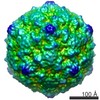 6638C M: map data used to model this data C: citing same article ( |
|---|---|
| Similar structure data |


-Links
 PDBj
PDBj


- Assembly
| Deposited unit | 
|
|---|---|
| 1 | x 60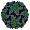
|
| 2 |
|
| 3 | x 5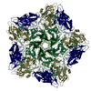
|
| 4 | x 6
|
| 5 | 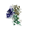
|
| Symmetry | Point symmetry: (Schoenflies symbol: I (icosahedral)) |
-Components
| #1: Protein | Mass: 31380.068 Da / Num. of mol.: 1 / Fragment: UNP residues 571-851 / Source method: isolated from a natural source / Source: (natural) Coxsackievirus B3 / References: UniProt: Q66282, UniProt: Q5UEA3*PLUS |
|---|---|
| #2: Protein | Mass: 28877.592 Da / Num. of mol.: 1 / Fragment: UNP residues 70-332 / Source method: isolated from a natural source / Source: (natural) Coxsackievirus B3 / References: UniProt: Q66282, UniProt: Q5UEA3*PLUS |
| #3: Protein | Mass: 26198.684 Da / Num. of mol.: 1 / Fragment: UNP residues 333-570 / Source method: isolated from a natural source / Source: (natural) Coxsackievirus B3 / References: UniProt: Q66282, UniProt: Q5UEA3*PLUS |
| #4: Protein | Mass: 7379.065 Da / Num. of mol.: 1 / Fragment: UNP residues 2-69 / Source method: isolated from a natural source / Source: (natural) Coxsackievirus B3 / References: UniProt: Q66282, UniProt: Q5UEA3*PLUS |
| #5: Chemical | ChemComp-PLM /   Mass: 256.424 Da / Num. of mol.: 1 / Source method: obtained synthetically / Formula: C16H32O2 Mass: 256.424 Da / Num. of mol.: 1 / Source method: obtained synthetically / Formula: C16H32O2 |
-Experimental details
-Experiment
| Experiment | Method: ELECTRON MICROSCOPY |
|---|---|
| EM experiment | Aggregation state: PARTICLE / 3D reconstruction method: single particle reconstruction |
- Sample preparation
| Component | Name: Human coxsackievirus B3 / Type: VIRUS |
|---|---|
| Details of virus | Empty: NO / Enveloped: NO / Host category: VERTEBRATES / Isolate: STRAIN / Type: VIRION |
| Specimen | Conc.: 1 mg/ml / Embedding applied: NO / Shadowing applied: NO / Staining applied: NO / Vitrification applied: YES |
| Vitrification | Instrument: GATAN CRYOPLUNGE 3 / Cryogen name: ETHANE / Temp: 102 K / Humidity: 90 % / Details: Plunged into liquid ethane (GATAN CRYOPLUNGE 3) |
- Electron microscopy imaging
| Experimental equipment |  Model: Tecnai Polara / Image courtesy: FEI Company |
|---|---|
| Microscopy | Model: FEI POLARA 300 / Date: Nov 21, 2014 |
| Electron gun | Electron source:  FIELD EMISSION GUN / Accelerating voltage: 300 kV / Illumination mode: FLOOD BEAM FIELD EMISSION GUN / Accelerating voltage: 300 kV / Illumination mode: FLOOD BEAM |
| Electron lens | Mode: BRIGHT FIELD / Nominal defocus max: 5875 nm / Nominal defocus min: 1362 nm / Cs: 2 mm |
| Specimen holder | Specimen holder model: SIDE ENTRY, EUCENTRIC |
| Image recording | Film or detector model: FEI FALCON II (4k x 4k) |
| Image scans | Num. digital images: 9685 |
- Processing
| CTF correction | Details: Each particle | ||||||||||||
|---|---|---|---|---|---|---|---|---|---|---|---|---|---|
| Symmetry | Point symmetry: I (icosahedral) | ||||||||||||
| 3D reconstruction | Resolution: 3.9 Å / Resolution method: FSC 0.143 / Num. of particles: 57203 / Nominal pixel size: 1.37 Å / Actual pixel size: 1.37 Å / Details: (Single particle--Applied symmetry: I) | ||||||||||||
| Atomic model building | Method: Real-space refinement / B value: 193 / Protocol: FLEXIBLE FIT / Space: REAL / Target criteria: Cross-correlation coefficient Details: Initial local fitting was done using Chimera. Phenix was then used for real-space refinement. | ||||||||||||
| Atomic model building | PDB-ID: 1COV Pdb chain-ID: 4 / Accession code: 1COV / Source name: PDB / Type: experimental model | ||||||||||||
| Refinement step | Cycle: LAST
|