National Institutes of Health/National Institute of General Medical Sciences (NIH/NIGMS)
P50GM103297
United States
National Institutes of Health/National Institute of General Medical Sciences (NIH/NIGMS)
R01GM080139
United States
National Institutes of Health/National Institute of General Medical Sciences (NIH/NIGMS)
P41GM103832
United States
National Institutes of Health/National Institute of General Medical Sciences (NIH/NIGMS)
R01GM42561
United States
Citation
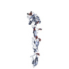
Journal: Structure / Year: 2018 Title: Structure of the 30 kDa HIV-1 RNA Dimerization Signal by a Hybrid Cryo-EM, NMR, and Molecular Dynamics Approach. Authors: Kaiming Zhang / Sarah C Keane / Zhaoming Su / Rossitza N Irobalieva / Muyuan Chen / Verna Van / Carly A Sciandra / Jan Marchant / Xiao Heng / Michael F Schmid / David A Case / Steven J ...Authors: Kaiming Zhang / Sarah C Keane / Zhaoming Su / Rossitza N Irobalieva / Muyuan Chen / Verna Van / Carly A Sciandra / Jan Marchant / Xiao Heng / Michael F Schmid / David A Case / Steven J Ludtke / Michael F Summers / Wah Chiu / Abstract: Cryoelectron microscopy (cryo-EM) and nuclear magnetic resonance (NMR) spectroscopy are routinely used to determine structures of macromolecules with molecular weights over 65 and under 25 kDa, ...Cryoelectron microscopy (cryo-EM) and nuclear magnetic resonance (NMR) spectroscopy are routinely used to determine structures of macromolecules with molecular weights over 65 and under 25 kDa, respectively. We combined these techniques to study a 30 kDa HIV-1 dimer initiation site RNA ([DIS]; 47 nt/strand). A 9 Å cryo-EM map clearly shows major groove features of the double helix and a right-handed superhelical twist. Simulated cryo-EM maps generated from time-averaged molecular dynamics trajectories (10 ns) exhibited levels of detail similar to those in the experimental maps, suggesting internal structural flexibility limits the cryo-EM resolution. Simultaneous inclusion of the cryo-EM map and H-edited NMR-derived distance restraints during structure refinement generates a structure consistent with both datasets and supporting a flipped-out base within a conserved purine-rich bulge. Our findings demonstrate the power of combining global and local structural information from these techniques for structure determination of modest-sized RNAs.
History
Deposition
Oct 20, 2017
-
Header (metadata) release
Nov 1, 2017
-
Map release
Feb 21, 2018
-
Update
Jan 29, 2020
-
Current status
Jan 29, 2020
Processing site: RCSB / Status: Released
-
Structure visualization
Movie
Surface view with section colored by density value
In the structure databanks used in Yorodumi, some data are registered as the other names, "COVID-19 virus" and "2019-nCoV". Here are the details of the virus and the list of structure data.
Jan 31, 2019. EMDB accession codes are about to change! (news from PDBe EMDB page)
EMDB accession codes are about to change! (news from PDBe EMDB page)
The allocation of 4 digits for EMDB accession codes will soon come to an end. Whilst these codes will remain in use, new EMDB accession codes will include an additional digit and will expand incrementally as the available range of codes is exhausted. The current 4-digit format prefixed with “EMD-” (i.e. EMD-XXXX) will advance to a 5-digit format (i.e. EMD-XXXXX), and so on. It is currently estimated that the 4-digit codes will be depleted around Spring 2019, at which point the 5-digit format will come into force.
The EM Navigator/Yorodumi systems omit the EMD- prefix.
Related info.:Q: What is EMD? / ID/Accession-code notation in Yorodumi/EM Navigator
Yorodumi is a browser for structure data from EMDB, PDB, SASBDB, etc.
This page is also the successor to EM Navigator detail page, and also detail information page/front-end page for Omokage search.
The word "yorodu" (or yorozu) is an old Japanese word meaning "ten thousand". "mi" (miru) is to see.
Related info.:EMDB / PDB / SASBDB / Comparison of 3 databanks / Yorodumi Search / Aug 31, 2016. New EM Navigator & Yorodumi / Yorodumi Papers / Jmol/JSmol / Function and homology information / Changes in new EM Navigator and Yorodumi
 Movie
Movie Controller

 Yorodumi
 Open data
 Basic information
 Map data
 Sample

 Human immunodeficiency virus 1
 Authors
 United States, 4 items
  Citation
 Structure visualization
 Movie viewer
 Downloads & links
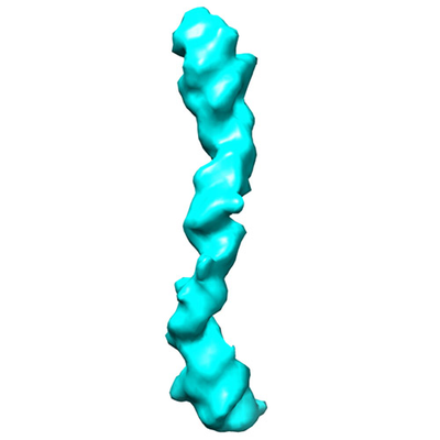 emd_7079.png
 http://ftp.pdbj.org/pub/emdb/structures/EMD-7079


 Sample components
 Processing
 Electron microscopy