 Movie
Movie Controller

[English] 日本語
 Yorodumi
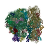
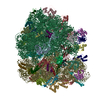
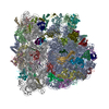
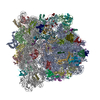
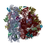
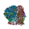
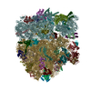
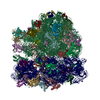
- PDB-5ngm: 2.9S structure of the 70S ribosome composing the S. aureus 100S c... -
+ Open data

- Basic information
| Entry | Database: PDB / ID: 5ngm | ||||||
|---|---|---|---|---|---|---|---|
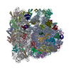
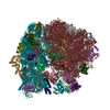
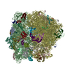
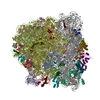
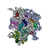
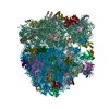
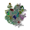
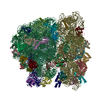
| Title | 2.9S structure of the 70S ribosome composing the S. aureus 100S complex | ||||||
 Components Components |
| ||||||
 Keywords Keywords | RIBOSOME / Ribosome Cryo-EM Structural Biology Hibernation | ||||||
| Function / homology |  Function and homology information Function and homology informationnegative regulation of translational elongation / ribosomal small subunit binding / large ribosomal subunit / ribosomal small subunit biogenesis / ribosomal small subunit assembly / small ribosomal subunit / small ribosomal subunit rRNA binding / transferase activity / 5S rRNA binding / large ribosomal subunit rRNA binding ...negative regulation of translational elongation / ribosomal small subunit binding / large ribosomal subunit / ribosomal small subunit biogenesis / ribosomal small subunit assembly / small ribosomal subunit / small ribosomal subunit rRNA binding / transferase activity / 5S rRNA binding / large ribosomal subunit rRNA binding / cytosolic small ribosomal subunit / cytosolic large ribosomal subunit / cytoplasmic translation / tRNA binding / rRNA binding / negative regulation of translation / ribosome / structural constituent of ribosome / translation / ribonucleoprotein complex / mRNA binding / zinc ion binding / cytoplasm / cytosol Similarity search - Function | ||||||
| Biological species |   Staphylococcus aureus (bacteria) Staphylococcus aureus (bacteria) | ||||||
| Method | ELECTRON MICROSCOPY / single particle reconstruction / cryo EM / Resolution: 2.9 Å | ||||||
 Authors Authors | Matzov, D. / Aibara, S. / Zimmerman, E. / Bashan, A. / Amunts, A. / Yonath, A. | ||||||
 Citation Citation | Journal: Nat Commun / Year: 2017 Title: The cryo-EM structure of hibernating 100S ribosome dimer from pathogenic Staphylococcus aureus. Authors: Donna Matzov / Shintaro Aibara / Arnab Basu / Ella Zimmerman / Anat Bashan / Mee-Ngan F Yap / Alexey Amunts / Ada E Yonath /    Abstract: Formation of 100S ribosome dimer is generally associated with translation suppression in bacteria. Trans-acting factors ribosome modulation factor (RMF) and hibernating promoting factor (HPF) were ...Formation of 100S ribosome dimer is generally associated with translation suppression in bacteria. Trans-acting factors ribosome modulation factor (RMF) and hibernating promoting factor (HPF) were shown to directly mediate this process in E. coli. Gram-positive S. aureus lacks an RMF homolog and the structural basis for its 100S formation was not known. Here we report the cryo-electron microscopy structure of the native 100S ribosome from S. aureus, revealing the molecular mechanism of its formation. The structure is distinct from previously reported analogs and relies on the HPF C-terminal extension forming the binding platform for the interactions between both of the small ribosomal subunits. The 100S dimer is formed through interactions between rRNA h26, h40, and protein uS2, involving conformational changes of the head as well as surface regions that could potentially prevent RNA polymerase from docking to the ribosome.Under conditions of nutrient limitation, bacterial ribosomes undergo dimerization, forming a 100S complex that is translationally inactive. Here the authors present the structural basis for formation of the 100S complexes in Gram-positive bacteria, shedding light on the mechanism of translation suppression by the ribosome-silencing factors. | ||||||
| History |
|
- Structure visualization
| Movie |
Movie viewer |
|---|---|
| Structure viewer | Molecule: MolmilJmol/JSmol |
- Downloads & links
-Download
| PDBx/mmCIF format | 5ngm.cif.gz | 3.1 MB | Display | PDBx/mmCIF format |
|---|---|---|---|---|
| PDB format | pdb5ngm.ent.gz | Display | PDB format | |
| PDBx/mmJSON format | 5ngm.json.gz | Tree view | PDBx/mmJSON format | |
| Others |  Other downloads Other downloads |
-Validation report
| Summary document | 5ngm_validation.pdf.gz | 1.4 MB | Display | wwPDB validaton report |
|---|---|---|---|---|
| Full document | 5ngm_full_validation.pdf.gz | 1.5 MB | Display | |
| Data in XML | 5ngm_validation.xml.gz | 204.4 KB | Display | |
| Data in CIF | 5ngm_validation.cif.gz | 355 KB | Display | |
| Arichive directory | https://data.pdbj.org/pub/pdb/validation_reports/ng/5ngmftp://data.pdbj.org/pub/pdb/validation_reports/ng/5ngm | HTTPS FTP |
-Related structure data
| Related structure data | 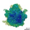 3640MC 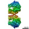 3637C 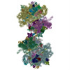 6fxcC C: citing same article ( M: map data used to model this data |
|---|---|
| Similar structure data |
-Links
 PDBj
PDBj


- Assembly
| Deposited unit | 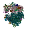
|
|---|---|
| 1 |
|
-Components
-RNA chain , 3 types, 3 molecules AaAAAB
| #1: RNA chain | Mass: 502975.906 Da / Num. of mol.: 1 / Source method: isolated from a natural source / Source: (natural) Staphylococcus aureus (bacteria) / References: GenBank: 1133392934 |
|---|---|
| #23: RNA chain | Mass: 946697.625 Da / Num. of mol.: 1 / Source method: isolated from a natural source / Source: (natural) Staphylococcus aureus (bacteria) / References: GenBank: 610395799 |
| #24: RNA chain | Mass: 36974.945 Da / Num. of mol.: 1 / Source method: isolated from a natural source / Source: (natural) Staphylococcus aureus (bacteria) / References: GenBank: 1130374198 |
-30S ribosomal protein ... , 20 types, 20 molecules AbAcAdAeAfAgAhAiAjAkAlAmAnAoApAqArAsAtAu
| #2: Protein | Mass: 29136.369 Da / Num. of mol.: 1 / Source method: isolated from a natural source / Source: (natural) Staphylococcus aureus (bacteria) / References: UniProt: A0A077UDU1 |
|---|---|
| #3: Protein | Mass: 24143.867 Da / Num. of mol.: 1 / Source method: isolated from a natural source / Source: (natural) Staphylococcus aureus (bacteria) / References: UniProt: A0A077UU26 |
| #4: Protein | Mass: 23051.416 Da / Num. of mol.: 1 / Source method: isolated from a natural source / Source: (natural) Staphylococcus aureus (bacteria) / References: UniProt: W8TVK2 |
| #5: Protein | Mass: 17770.512 Da / Num. of mol.: 1 / Source method: isolated from a natural source / Source: (natural) Staphylococcus aureus (bacteria) / References: UniProt: W8TUC9 |
| #6: Protein | Mass: 11613.146 Da / Num. of mol.: 1 / Source method: isolated from a natural source / Source: (natural) Staphylococcus aureus (bacteria) / References: UniProt: W8TPC6 |
| #7: Protein | Mass: 17826.555 Da / Num. of mol.: 1 / Source method: isolated from a natural source / Source: (natural) Staphylococcus aureus (bacteria) / References: UniProt: A0A077VHU6 |
| #8: Protein | Mass: 14854.315 Da / Num. of mol.: 1 / Source method: isolated from a natural source / Source: (natural) Staphylococcus aureus (bacteria) / References: UniProt: W8U8T8 |
| #9: Protein | Mass: 14856.987 Da / Num. of mol.: 1 / Source method: isolated from a natural source / Source: (natural) Staphylococcus aureus (bacteria) / References: UniProt: A0A0K7UQP6 |
| #10: Protein | Mass: 11598.503 Da / Num. of mol.: 1 / Source method: isolated from a natural source / Source: (natural) Staphylococcus aureus (bacteria) / References: UniProt: A0A0H2K0A0 |
| #11: Protein | Mass: 13907.978 Da / Num. of mol.: 1 / Source method: isolated from a natural source / Source: (natural) Staphylococcus aureus (bacteria) / References: UniProt: A0A077UKD6 |
| #12: Protein | Mass: 15320.870 Da / Num. of mol.: 1 / Source method: isolated from a natural source / Source: (natural) Staphylococcus aureus (bacteria) / References: UniProt: W8U1C6 |
| #13: Protein | Mass: 13747.919 Da / Num. of mol.: 1 / Source method: isolated from a natural source / Source: (natural) Staphylococcus aureus (bacteria) / References: UniProt: W8U8U6 |
| #14: Protein | Mass: 7317.769 Da / Num. of mol.: 1 / Source method: isolated from a natural source / Source: (natural) Staphylococcus aureus (bacteria) / References: UniProt: A0A077UGW7 |
| #15: Protein | Mass: 10634.330 Da / Num. of mol.: 1 / Source method: isolated from a natural source / Source: (natural) Staphylococcus aureus (bacteria) / References: UniProt: W8U6H8 |
| #16: Protein | Mass: 10253.886 Da / Num. of mol.: 1 / Source method: isolated from a natural source / Source: (natural) Staphylococcus aureus (bacteria) / References: UniProt: W8U6K4 |
| #17: Protein | Mass: 10196.888 Da / Num. of mol.: 1 / Source method: isolated from a natural source / Source: (natural) Staphylococcus aureus (bacteria) / References: UniProt: A0A077VLD5 |
| #18: Protein | Mass: 9332.018 Da / Num. of mol.: 1 / Source method: isolated from a natural source / Source: (natural) Staphylococcus aureus (bacteria) / References: UniProt: A0A077UZF4 |
| #19: Protein | Mass: 10639.309 Da / Num. of mol.: 1 / Source method: isolated from a natural source / Source: (natural) Staphylococcus aureus (bacteria) / References: UniProt: A0A077V0P8 |
| #20: Protein | Mass: 9039.472 Da / Num. of mol.: 1 / Source method: isolated from a natural source / Source: (natural) Staphylococcus aureus (bacteria) / References: UniProt: W8UXC3 |
| #21: Protein | Mass: 6994.267 Da / Num. of mol.: 1 / Source method: isolated from a natural source / Source: (natural) Staphylococcus aureus (bacteria) / References: UniProt: A0A077UHS1 |
+50S ribosomal protein ... , 28 types, 28 molecules ACADAEAFAGAHAIAJAKALAMANAOAPAQARASATAUAVAWAXAYAZA1A2A3A4
-Protein / Non-polymers , 2 types, 206 molecules Av
| #22: Protein | Mass: 22244.914 Da / Num. of mol.: 1 / Source method: isolated from a natural source / Source: (natural) Staphylococcus aureus (bacteria) / References: UniProt: W8USK0, UniProt: D2Z097*PLUS |
|---|---|
| #53: Chemical | ChemComp-MG /  Mass: 24.305 Da / Num. of mol.: 205 / Source method: obtained synthetically / Formula: Mg Mass: 24.305 Da / Num. of mol.: 205 / Source method: obtained synthetically / Formula: Mg |
-Experimental details
-Experiment
| Experiment | Method: ELECTRON MICROSCOPY |
|---|---|
| EM experiment | Aggregation state: PARTICLE / 3D reconstruction method: single particle reconstruction |
- Sample preparation
| Component |
| ||||||||||||||||||||||||||||||||||||||||||||||||
|---|---|---|---|---|---|---|---|---|---|---|---|---|---|---|---|---|---|---|---|---|---|---|---|---|---|---|---|---|---|---|---|---|---|---|---|---|---|---|---|---|---|---|---|---|---|---|---|---|---|
| Molecular weight |
| ||||||||||||||||||||||||||||||||||||||||||||||||
| Source (natural) |
| ||||||||||||||||||||||||||||||||||||||||||||||||
| Buffer solution | pH: 7.5 | ||||||||||||||||||||||||||||||||||||||||||||||||
| Specimen | Embedding applied: NO / Shadowing applied: NO / Staining applied: NO / Vitrification applied: YES | ||||||||||||||||||||||||||||||||||||||||||||||||
| Vitrification | Cryogen name: ETHANE |
- Electron microscopy imaging
| Experimental equipment |  Model: Titan Krios / Image courtesy: FEI Company |
|---|---|
| Microscopy | Model: FEI TITAN KRIOS |
| Electron gun | Electron source:  FIELD EMISSION GUN / Accelerating voltage: 300 kV / Illumination mode: FLOOD BEAM FIELD EMISSION GUN / Accelerating voltage: 300 kV / Illumination mode: FLOOD BEAM |
| Electron lens | Mode: BRIGHT FIELD |
| Image recording | Electron dose: 2.3 e/Å2 / Film or detector model: FEI FALCON II (4k x 4k) |
- Processing
| CTF correction | Type: NONE |
|---|---|
| 3D reconstruction | Resolution: 2.9 Å / Resolution method: FSC 0.143 CUT-OFF / Num. of particles: 224554 / Symmetry type: POINT |
