 Movie
Movie Controller

[English] 日本語
 Yorodumi
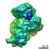
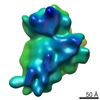
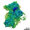
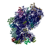
- EMDB-4071: Structure of the 40S ABCE1 post-splitting complex in ribosome rec... -
+ Open data

- Basic information
| Entry | Database: EMDB / ID: EMD-4071 | |||||||||
|---|---|---|---|---|---|---|---|---|---|---|
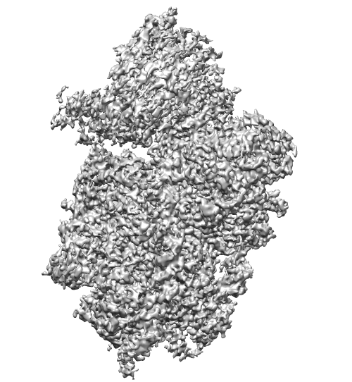
| Title | Structure of the 40S ABCE1 post-splitting complex in ribosome recycling and translation initiation | |||||||||
 Map data Map data | ||||||||||
 Sample Sample |
| |||||||||
| Function / homology |  Function and homology information Function and homology informationribosome disassembly /  Protein methylation / RMTs methylate histone arginines / positive regulation of translational fidelity / mTORC1-mediated signalling / Protein hydroxylation / : / Formation of the ternary complex, and subsequently, the 43S complex / Translation initiation complex formation / Ribosomal scanning and start codon recognition ...ribosome disassembly / Protein methylation / RMTs methylate histone arginines / positive regulation of translational fidelity / mTORC1-mediated signalling / Protein hydroxylation / : / Formation of the ternary complex, and subsequently, the 43S complex / Translation initiation complex formation / Ribosomal scanning and start codon recognition / ribosomal subunit export from nucleus / Major pathway of rRNA processing in the nucleolus and cytosol / SRP-dependent cotranslational protein targeting to membrane / 90S preribosome / GTP hydrolysis and joining of the 60S ribosomal subunit / Formation of a pool of free 40S subunits / endonucleolytic cleavage to generate mature 3'-end of SSU-rRNA from (SSU-rRNA, 5.8S rRNA, LSU-rRNA) / Nonsense Mediated Decay (NMD) independent of the Exon Junction Complex (EJC) / Nonsense Mediated Decay (NMD) enhanced by the Exon Junction Complex (EJC) / ribosomal small subunit binding / L13a-mediated translational silencing of Ceruloplasmin expression / endonucleolytic cleavage in ITS1 to separate SSU-rRNA from 5.8S rRNA and LSU-rRNA from tricistronic rRNA transcript (SSU-rRNA, 5.8S rRNA, LSU-rRNA) / regulation of translational fidelity / translational termination / maturation of SSU-rRNA from tricistronic rRNA transcript (SSU-rRNA, 5.8S rRNA, LSU-rRNA) / ribosomal large subunit biogenesis / translational initiation / ribosome assembly / translation initiation factor activity / small-subunit processome / positive regulation of translation / maintenance of translational fidelity / ribosomal small subunit biogenesis / small ribosomal subunit rRNA binding / ribosomal small subunit assembly / rRNA processing / cytoplasmic stress granule / cytosolic small ribosomal subunit / ribosome biogenesis / cytoplasmic translation / rRNA binding / ribosome / structural constituent of ribosome / translation / iron ion binding / mRNA binding / nucleolus / ATP hydrolysis activity / mitochondrion / RNA binding / nucleoplasm / ATP binding / metal ion binding / nucleus / cytosol / cytoplasm Protein methylation / RMTs methylate histone arginines / positive regulation of translational fidelity / mTORC1-mediated signalling / Protein hydroxylation / : / Formation of the ternary complex, and subsequently, the 43S complex / Translation initiation complex formation / Ribosomal scanning and start codon recognition ...ribosome disassembly / Protein methylation / RMTs methylate histone arginines / positive regulation of translational fidelity / mTORC1-mediated signalling / Protein hydroxylation / : / Formation of the ternary complex, and subsequently, the 43S complex / Translation initiation complex formation / Ribosomal scanning and start codon recognition / ribosomal subunit export from nucleus / Major pathway of rRNA processing in the nucleolus and cytosol / SRP-dependent cotranslational protein targeting to membrane / 90S preribosome / GTP hydrolysis and joining of the 60S ribosomal subunit / Formation of a pool of free 40S subunits / endonucleolytic cleavage to generate mature 3'-end of SSU-rRNA from (SSU-rRNA, 5.8S rRNA, LSU-rRNA) / Nonsense Mediated Decay (NMD) independent of the Exon Junction Complex (EJC) / Nonsense Mediated Decay (NMD) enhanced by the Exon Junction Complex (EJC) / ribosomal small subunit binding / L13a-mediated translational silencing of Ceruloplasmin expression / endonucleolytic cleavage in ITS1 to separate SSU-rRNA from 5.8S rRNA and LSU-rRNA from tricistronic rRNA transcript (SSU-rRNA, 5.8S rRNA, LSU-rRNA) / regulation of translational fidelity / translational termination / maturation of SSU-rRNA from tricistronic rRNA transcript (SSU-rRNA, 5.8S rRNA, LSU-rRNA) / ribosomal large subunit biogenesis / translational initiation / ribosome assembly / translation initiation factor activity / small-subunit processome / positive regulation of translation / maintenance of translational fidelity / ribosomal small subunit biogenesis / small ribosomal subunit rRNA binding / ribosomal small subunit assembly / rRNA processing / cytoplasmic stress granule / cytosolic small ribosomal subunit / ribosome biogenesis / cytoplasmic translation / rRNA binding / ribosome / structural constituent of ribosome / translation / iron ion binding / mRNA binding / nucleolus / ATP hydrolysis activity / mitochondrion / RNA binding / nucleoplasm / ATP binding / metal ion binding / nucleus / cytosol / cytoplasmSimilarity search - Function | |||||||||
| Biological species |  Saccharomyces cerevisiae (brewer's yeast) Saccharomyces cerevisiae (brewer's yeast) | |||||||||
| Method | single particle reconstruction / cryo EM / Resolution: 3.9 Å | |||||||||
 Authors Authors | Heuer A / Gerovac M / Schmidt C / Trowitzsch S / Preis A / Koetter P / Berninghausen O / Becker T / Beckmann R / Tampe R | |||||||||
 Citation Citation | Journal: Nat Struct Mol Biol / Year: 2017 Title: Structure of the 40S-ABCE1 post-splitting complex in ribosome recycling and translation initiation. Authors: André Heuer / Milan Gerovac / Christian Schmidt / Simon Trowitzsch / Anne Preis / Peter Kötter / Otto Berninghausen / Thomas Becker / Roland Beckmann / Robert Tampé /  Abstract: The essential ATP-binding cassette protein ABCE1 splits 80S ribosomes into 60S and 40S subunits after canonical termination or quality-control-based mRNA surveillance processes. However, the ...The essential ATP-binding cassette protein ABCE1 splits 80S ribosomes into 60S and 40S subunits after canonical termination or quality-control-based mRNA surveillance processes. However, the underlying splitting mechanism remains enigmatic. Here, we present a cryo-EM structure of the yeast 40S-ABCE1 post-splitting complex at 3.9-Å resolution. Compared to the pre-splitting state, we observe repositioning of ABCE1's iron-sulfur cluster domain, which rotates 150° into a binding pocket on the 40S subunit. This repositioning explains a newly observed anti-association activity of ABCE1. Notably, the movement implies a collision with A-site factors, thus explaining the splitting mechanism. Disruption of key interactions in the post-splitting complex impairs cellular homeostasis. Additionally, the structure of a native post-splitting complex reveals ABCE1 to be part of the 43S initiation complex, suggesting a coordination of termination, recycling, and initiation. | |||||||||
| History |
|
- Structure visualization
| Movie |
Movie viewer |
|---|---|
| Structure viewer | EM map: SurfViewMolmilJmol/JSmol |
| Supplemental images |

- Downloads & links
-EMDB archive
| Map data | emd_4071.map.gz | 202.2 MB | EMDB map data format | |
|---|---|---|---|---|
| Header (meta data) | emd-4071-v30.xmlemd-4071.xml | 12.1 KB 12.1 KB | Display Display | EMDB header |
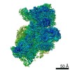
| Images |  emd_4071.jpg emd_4071.jpg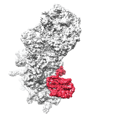 emd_4071.png emd_4071.png | 214.9 KB 153.6 KB | ||
| Archive directory |  http://ftp.pdbj.org/pub/emdb/structures/EMD-4071ftp://ftp.pdbj.org/pub/emdb/structures/EMD-4071 http://ftp.pdbj.org/pub/emdb/structures/EMD-4071ftp://ftp.pdbj.org/pub/emdb/structures/EMD-4071 | HTTPS FTP |
-Related structure data
| Related structure data |  5ll6MC  3452C M: atomic model generated by this map C: citing same article ( |
|---|---|
| Similar structure data |


-Links
| EMDB pages | EMDB (EBI/PDBe) / EMDataResource |
|---|---|
| Related items in Molecule of the Month |


-Map
| File | Download / File: emd_4071.map.gz / Format: CCP4 / Size: 216 MB / Type: IMAGE STORED AS FLOATING POINT NUMBER (4 BYTES) | ||||||||||||||||||||||||||||||||||||||||||||||||||||||||||||
|---|---|---|---|---|---|---|---|---|---|---|---|---|---|---|---|---|---|---|---|---|---|---|---|---|---|---|---|---|---|---|---|---|---|---|---|---|---|---|---|---|---|---|---|---|---|---|---|---|---|---|---|---|---|---|---|---|---|---|---|---|---|
| Voxel size | X=Y=Z: 1.084 Å | ||||||||||||||||||||||||||||||||||||||||||||||||||||||||||||
| Density |
| ||||||||||||||||||||||||||||||||||||||||||||||||||||||||||||
| Symmetry | Space group: 1 | ||||||||||||||||||||||||||||||||||||||||||||||||||||||||||||
| Details | EMDB XML:
CCP4 map header:
| ||||||||||||||||||||||||||||||||||||||||||||||||||||||||||||
-Supplemental data
- Sample components
-Entire : 40S-ABCE1 complex
| Entire | Name: 40S-ABCE1 complex |
|---|---|
| Components |
|
-Supramolecule #1: 40S-ABCE1 complex
| Supramolecule | Name: 40S-ABCE1 complex / type: complex / ID: 1 / Parent: 0 |
|---|---|
| Source (natural) | Organism: Saccharomyces cerevisiae (brewer's yeast) |
| Molecular weight | Theoretical: 1.2 MDa |
-Experimental details
-Structure determination
| Method | cryo EM |
|---|---|
 Processing Processing | single particle reconstruction |
| Aggregation state | particle |
-Sample preparation
| Buffer | pH: 7.4 Component:
| |||||||||||||||
|---|---|---|---|---|---|---|---|---|---|---|---|---|---|---|---|---|
| Grid | Model: Quantifoil R3/3 / Material: COPPER/PALLADIUM / Support film - Material: CARBON / Support film - topology: CONTINUOUS / Support film - Film thickness: 2.0 nm / Pretreatment - Type: GLOW DISCHARGE | |||||||||||||||
| Vitrification | Cryogen name: ETHANE / Chamber humidity: 100 % / Chamber temperature: 278 K / Instrument: FEI VITROBOT MARK IV | |||||||||||||||
| Details | Purified 40S ribosomal subunits were reconstituted with purified, recombinantly expressed ABCE1 |
- Electron microscopy
| Microscope | FEI TITAN |
|---|---|
| Electron beam | Acceleration voltage: 300 kV / Electron source: FIELD EMISSION GUN |
| Electron optics | Calibrated defocus min: 0.8 µm / Illumination mode: FLOOD BEAM / Imaging mode: BRIGHT FIELDBright-field microscopy / Cs: 2.7 mm / Nominal defocus max: 2.4 µm |
| Sample stage | Specimen holder model: FEI TITAN KRIOS AUTOGRID HOLDER / Cooling holder cryogen: NITROGEN |
| Image recording | Film or detector model: FEI FALCON II (4k x 4k) / Average electron dose: 2.4 e/Å2 |
-Image processing
| CTF correction | Software - Name: CTFFIND (ver. 4) |
|---|---|
| Startup model | Type of model: OTHER / Details: yeast 40S ribosome |
| Initial angle assignment | Type: PROJECTION MATCHING / Software - Name: RELION (ver. 2.0) |
| Final 3D classification | Software - Name: RELION (ver. 2.0) |
| Final angle assignment | Type: PROJECTION MATCHING / Software - Name: RELION (ver. 2.0) |
| Final reconstruction | Resolution.type: BY AUTHOR / Resolution: 3.9 Å / Resolution method: FSC 0.143 CUT-OFF / Software - Name: RELION (ver. 2.0) / Number images used: 102000 |
-Atomic model buiding 1
| Refinement | Space: REAL / Protocol: FLEXIBLE FIT |
|---|---|
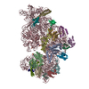
| Output model | PDB-5ll6: |
