 Movie
Movie Controller

[English] 日本語
 Yorodumi
- EMDB-8417: Time-resolved cryo electron microscopy map of the tRNA-bound 30S ... -
+ Open data

- Basic information
| Entry | Database: EMDB / ID: EMD-8417 | |||||||||||||||
|---|---|---|---|---|---|---|---|---|---|---|---|---|---|---|---|---|
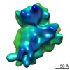
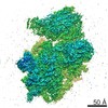
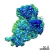
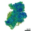
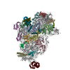
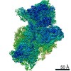
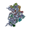
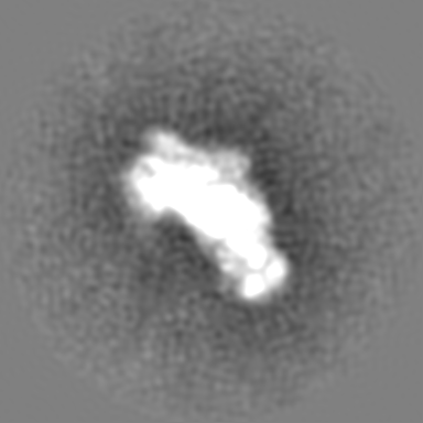
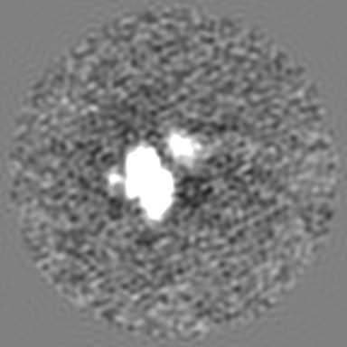
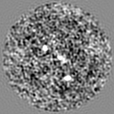
| Title | Time-resolved cryo electron microscopy map of the tRNA-bound 30S subunit | |||||||||||||||
 Map data Map data | tRNA bound 30S subunit | |||||||||||||||
 Sample Sample |
| |||||||||||||||
| Biological species |  | |||||||||||||||
| Method | single particle reconstruction / cryo EM / Resolution: 10.0 Å | |||||||||||||||
 Authors Authors | Fu Z / Kaledhonkar S / Borg A / Sun M / Chen B / Grassucci RA / Ehrenberg M / Frank J | |||||||||||||||
| Funding support |  United States, United States,  Sweden, 4 items Sweden, 4 items
| |||||||||||||||
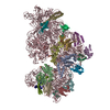
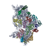
 Citation Citation | Journal: Structure / Year: 2016 Title: Key Intermediates in Ribosome Recycling Visualized by Time-Resolved Cryoelectron Microscopy. Authors: Ziao Fu / Sandip Kaledhonkar / Anneli Borg / Ming Sun / Bo Chen / Robert A Grassucci / Måns Ehrenberg / Joachim Frank / Abstract: Upon encountering a stop codon on mRNA, polypeptide synthesis on the ribosome is terminated by release factors, and the ribosome complex, still bound with mRNA and P-site-bound tRNA (post-termination ...Upon encountering a stop codon on mRNA, polypeptide synthesis on the ribosome is terminated by release factors, and the ribosome complex, still bound with mRNA and P-site-bound tRNA (post-termination complex, PostTC), is split into ribosomal subunits, ready for a new round of translational initiation. Separation of post-termination ribosomes into subunits, or "ribosome recycling," is promoted by the joint action of ribosome-recycling factor (RRF) and elongation factor G (EF-G) in a guanosine triphosphate (GTP) hydrolysis-dependent manner. Here we used a mixing-spraying-based method of time-resolved cryo-electron microscopy (cryo-EM) to visualize the short-lived intermediates of the recycling process. The two complexes that contain (1) both RRF and EF-G bound to the PostTC or (2) deacylated tRNA bound to the 30S subunit are of particular interest. Our observations of the native form of these complexes demonstrate the strong potential of time-resolved cryo-EM for visualizing previously unobservable transient structures. | |||||||||||||||
| History |
|
- Structure visualization
| Movie |
 Movie viewer Movie viewer |
|---|---|
| Structure viewer | EM map: SurfViewMolmilJmol/JSmol |
| Supplemental images |

- Downloads & links
-EMDB archive
| Map data | emd_8417.map.gz | 201.6 MB | EMDB map data format | |
|---|---|---|---|---|
| Header (meta data) | emd-8417-v30.xmlemd-8417.xml | 14.1 KB 14.1 KB | Display Display | EMDB header |
| Images | 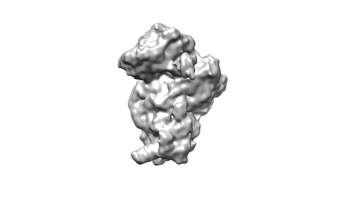 emd_8417.png emd_8417.png | 38.9 KB | ||
| Archive directory |  http://ftp.pdbj.org/pub/emdb/structures/EMD-8417ftp://ftp.pdbj.org/pub/emdb/structures/EMD-8417 http://ftp.pdbj.org/pub/emdb/structures/EMD-8417ftp://ftp.pdbj.org/pub/emdb/structures/EMD-8417 | HTTPS FTP |
-Related structure data
| Related structure data |  8411C  8412C  8413C 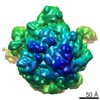 8415C 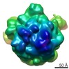 8416C 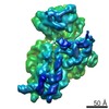 8418C C: citing same article ( |
|---|---|
| Similar structure data |

-Links
| EMDB pages | EMDB (EBI/PDBe) / EMDataResource |
|---|---|
| Related items in Molecule of the Month |

-Map
| File | Download / File: emd_8417.map.gz / Format: CCP4 / Size: 216 MB / Type: IMAGE STORED AS FLOATING POINT NUMBER (4 BYTES) | ||||||||||||||||||||||||||||||||||||||||||||||||||||||||||||||||||||
|---|---|---|---|---|---|---|---|---|---|---|---|---|---|---|---|---|---|---|---|---|---|---|---|---|---|---|---|---|---|---|---|---|---|---|---|---|---|---|---|---|---|---|---|---|---|---|---|---|---|---|---|---|---|---|---|---|---|---|---|---|---|---|---|---|---|---|---|---|---|
| Annotation | tRNA bound 30S subunit | ||||||||||||||||||||||||||||||||||||||||||||||||||||||||||||||||||||
| Projections & slices | Image control
Images are generated by Spider. | ||||||||||||||||||||||||||||||||||||||||||||||||||||||||||||||||||||
| Voxel size | X=Y=Z: 1.25 Å | ||||||||||||||||||||||||||||||||||||||||||||||||||||||||||||||||||||
| Density |
| ||||||||||||||||||||||||||||||||||||||||||||||||||||||||||||||||||||
| Symmetry | Space group: 1 | ||||||||||||||||||||||||||||||||||||||||||||||||||||||||||||||||||||
| Details | EMDB XML:
CCP4 map header:
| ||||||||||||||||||||||||||||||||||||||||||||||||||||||||||||||||||||
 Z (Sec.)
 Y (Row.)
 X (Col.)


-Supplemental data
- Sample components
-Entire : tRNA-bound 30S subunit
| Entire | Name: tRNA-bound 30S subunit |
|---|---|
| Components |
|
-Supramolecule #1: tRNA-bound 30S subunit
| Supramolecule | Name: tRNA-bound 30S subunit / type: complex / ID: 1 / Parent: 0 |
|---|
-Supramolecule #2: tRNA
| Supramolecule | Name: tRNA / type: complex / ID: 2 / Parent: 1 |
|---|---|
| Source (natural) | Organism: |
| Recombinant expression | Organism: |
-Supramolecule #3: 30S ribosomal subunit
| Supramolecule | Name: 30S ribosomal subunit / type: complex / ID: 3 / Parent: 1 |
|---|---|
| Source (natural) | Organism: |
-Supramolecule #4: mRNA
| Supramolecule | Name: mRNA / type: complex / ID: 4 / Parent: 1 Details: Encodes peptide fMet-Phe-Thr (sequence GGGAAUUCGGGCCCUUGUUAACAAUUAAGGAGGUAUUAAAUGUUUACGUAAUUGCAGAAAAAAAAAAAAAAAAAAAAA) |
|---|---|
| Source (natural) | Organism: |
| Recombinant expression | Organism: |
-Experimental details
-Structure determination
| Method | cryo EM |
|---|---|
 Processing Processing | single particle reconstruction |
| Aggregation state | particle |
-Sample preparation
| Concentration | 2.7 mg/mL |
|---|---|
| Buffer | pH: 7.5 |
| Grid | Model: Quantifoil R1.2/1.3 / Material: COPPER / Mesh: 300 |
| Vitrification | Cryogen name: ETHANE / Chamber humidity: 90 % / Chamber temperature: 298 K / Instrument: HOMEMADE PLUNGER |
- Electron microscopy
| Microscope | FEI TECNAI F30 |
|---|---|
| Temperature | Min: 80.0 K |
| Specialist optics | Spherical aberration corrector: None / Chromatic aberration corrector: None / Energy filter - Name: None |
| Image recording | Film or detector model: GATAN K2 SUMMIT (4k x 4k) / Detector mode: COUNTING / Digitization - Dimensions - Width: 3710 pixel / Digitization - Dimensions - Height: 3838 pixel / Digitization - Sampling interval: 5.0 µm / Digitization - Frames/image: 1-50 / Average exposure time: 0.2 sec. / Average electron dose: 1.01 e/Å2 |
| Electron beam | Acceleration voltage: 300 kV / Electron source:  FIELD EMISSION GUN FIELD EMISSION GUN |
| Electron optics | C2 aperture diameter: 30.0 µm / Illumination mode: FLOOD BEAM / Imaging mode: BRIGHT FIELD / Cs: 2.26 mm / Nominal defocus max: 3.0 µm / Nominal defocus min: 1.5 µm / Nominal magnification: 31000 |
| Sample stage | Specimen holder model: SIDE ENTRY, EUCENTRIC / Cooling holder cryogen: NITROGEN |
| Experimental equipment |  Model: Tecnai F30 / Image courtesy: FEI Company |
-Image processing
| CTF correction | Software - Name: CTFFIND3 |
|---|---|
| Final reconstruction | Resolution.type: BY AUTHOR / Resolution: 10.0 Å / Resolution method: FSC 0.143 CUT-OFF / Number images used: 5241 |
| Initial angle assignment | Type: PROJECTION MATCHING / Software - Name: RELION (ver. 1.3) |
| Final angle assignment | Type: ANGULAR RECONSTITUTION / Software - Name: RELION (ver. 1.3) |
