ムービー
ムービー コントローラー


+ データを開く

- 基本情報
基本情報
| 登録情報 | データベース: EMDB / ID: EMD-9512 | ||||||||||||
|---|---|---|---|---|---|---|---|---|---|---|---|---|---|
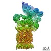
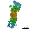
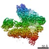
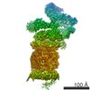
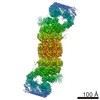
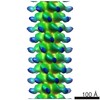
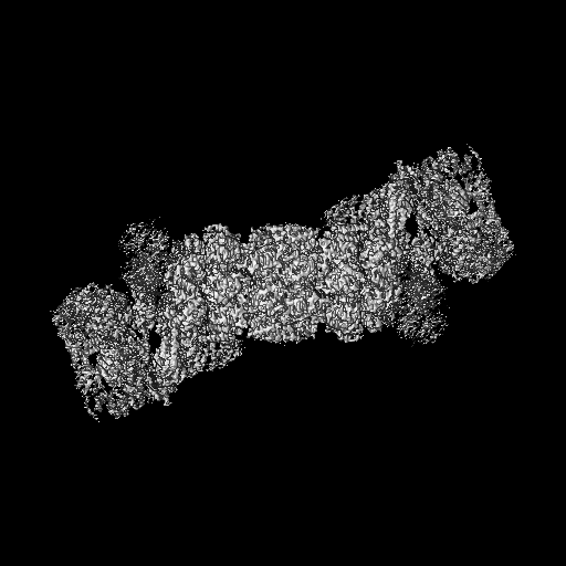
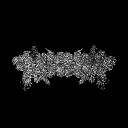
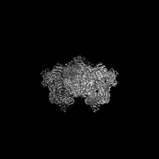
| タイトル | Cryo-EM map of the human 26S proteasome at 3.5A resolution with C2 symmetry | ||||||||||||
 マップデータ マップデータ | with C2 symmetry | ||||||||||||
 試料 試料 |
| ||||||||||||
 キーワード キーワード | protein complex / human proteasome / HYDROLASE | ||||||||||||
| 機能・相同性 |  機能・相同性情報 機能・相同性情報thyrotropin-releasing hormone receptor binding / Impaired BRCA2 translocation to the nucleus / Impaired BRCA2 binding to SEM1 (DSS1) / nuclear proteasome complex / host-mediated perturbation of viral transcription / positive regulation of inclusion body assembly / integrator complex / proteasome accessory complex / meiosis I / purine ribonucleoside triphosphate binding ...thyrotropin-releasing hormone receptor binding / Impaired BRCA2 translocation to the nucleus / Impaired BRCA2 binding to SEM1 (DSS1) / nuclear proteasome complex / host-mediated perturbation of viral transcription / positive regulation of inclusion body assembly / integrator complex / proteasome accessory complex / meiosis I / purine ribonucleoside triphosphate binding / proteasome regulatory particle / cytosolic proteasome complex / positive regulation of proteasomal protein catabolic process / proteasome-activating activity / Antigen processing: Ub, ATP-independent proteasomal degradation / proteasome regulatory particle, lid subcomplex / proteasome regulatory particle, base subcomplex / sperm glycocalyx / protein K63-linked deubiquitination / negative regulation of programmed cell death / metal-dependent deubiquitinase activity / Regulation of ornithine decarboxylase (ODC) / Proteasome assembly / proteasome core complex / perinuclear theca / Cross-presentation of soluble exogenous antigens (endosomes) / Somitogenesis / K63-linked deubiquitinase activity / transcription factor binding / Homologous DNA Pairing and Strand Exchange / Defective homologous recombination repair (HRR) due to BRCA1 loss of function / Defective HDR through Homologous Recombination Repair (HRR) due to PALB2 loss of BRCA1 binding function / Defective HDR through Homologous Recombination Repair (HRR) due to PALB2 loss of BRCA2/RAD51/RAD51C binding function / Resolution of D-loop Structures through Synthesis-Dependent Strand Annealing (SDSA) / Resolution of D-loop Structures through Holliday Junction Intermediates / proteasome binding / Impaired BRCA2 binding to RAD51 / myofibril / regulation of protein catabolic process / proteasome storage granule / proteasomal ubiquitin-independent protein catabolic process / sperm head-tail coupling apparatus / Presynaptic phase of homologous DNA pairing and strand exchange / general transcription initiation factor binding / protein deubiquitination / positive regulation of RNA polymerase II transcription preinitiation complex assembly / blastocyst development / polyubiquitin modification-dependent protein binding / immune system process / proteasome endopeptidase complex / NF-kappaB binding / proteasome core complex, beta-subunit complex / endopeptidase activator activity / threonine-type endopeptidase activity / proteasome assembly / mRNA export from nucleus / proteasome core complex, alpha-subunit complex / SARS-CoV-1 targets host intracellular signalling and regulatory pathways / enzyme regulator activity / ERAD pathway / regulation of proteasomal protein catabolic process / inclusion body / ciliary tip / : / TBP-class protein binding / proteasome complex / stem cell differentiation / Regulation of activated PAK-2p34 by proteasome mediated degradation / sarcomere / Autodegradation of Cdh1 by Cdh1:APC/C / APC/C:Cdc20 mediated degradation of Securin / ubiquitin binding / N-glycan trimming in the ER and Calnexin/Calreticulin cycle / Asymmetric localization of PCP proteins / Ubiquitin-dependent degradation of Cyclin D / SCF-beta-TrCP mediated degradation of Emi1 / NIK-->noncanonical NF-kB signaling / TNFR2 non-canonical NF-kB pathway / AUF1 (hnRNP D0) binds and destabilizes mRNA / centriole / sperm end piece / P-body / negative regulation of inflammatory response to antigenic stimulus / Assembly of the pre-replicative complex / Vpu mediated degradation of CD4 / Cdc20:Phospho-APC/C mediated degradation of Cyclin A / lipopolysaccharide binding / Degradation of DVL / Dectin-1 mediated noncanonical NF-kB signaling / Degradation of CRY and PER proteins / Degradation of AXIN / Hh mutants are degraded by ERAD / Activation of NF-kappaB in B cells / G2/M Checkpoints / Hedgehog ligand biogenesis / Degradation of GLI1 by the proteasome / Defective CFTR causes cystic fibrosis / Autodegradation of the E3 ubiquitin ligase COP1 / Regulation of RUNX3 expression and activity / GSK3B and BTRC:CUL1-mediated-degradation of NFE2L2 類似検索 - 分子機能 | ||||||||||||
| 生物種 |  Homo sapiens (ヒト) Homo sapiens (ヒト) | ||||||||||||
| 手法 | 単粒子再構成法 / クライオ電子顕微鏡法 / 解像度: 3.5 Å | ||||||||||||
 データ登録者 データ登録者 | Huang XL / Luan B / Wu JP / Shi YG | ||||||||||||
| 資金援助 |  中国, 3件 中国, 3件
| ||||||||||||
 引用 引用 | ジャーナル: Nat Struct Mol Biol / 年: 2016 タイトル: An atomic structure of the human 26S proteasome. 著者: Xiuliang Huang / Bai Luan / Jianping Wu / Yigong Shi / 要旨: We report the cryo-EM structure of the human 26S proteasome at an average resolution of 3.5 Å, allowing atomic modeling of 28 subunits in the core particle (CP) and 18 subunits in the regulatory ...We report the cryo-EM structure of the human 26S proteasome at an average resolution of 3.5 Å, allowing atomic modeling of 28 subunits in the core particle (CP) and 18 subunits in the regulatory particle (RP). The C-terminal residues of Rpt3 and Rpt5 subunits in the RP can be seen inserted into surface pockets formed between adjacent α subunits in the CP. Each of the six Rpt subunits contains a bound nucleotide, and the central gate of the CP α-ring is closed despite RP association. The six pore 1 loops in the Rpt ring are arranged similarly to a spiral staircase along the axial channel of substrate transport, which is constricted by the pore 2 loops. We also determined the cryo-EM structure of the human proteasome bound to the deubiquitinating enzyme USP14 at 4.35-Å resolution. Together, our structures provide a framework for mechanistic understanding of eukaryotic proteasome function. | ||||||||||||
| 履歴 |
|
- 構造の表示
構造の表示
| ムービー |
ムービービューア |
|---|---|
| 構造ビューア | EMマップ: SurfViewMolmilJmol/JSmol |
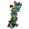
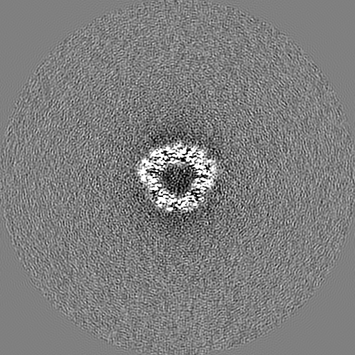
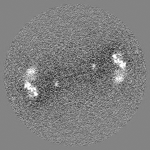
| 添付画像 |

- ダウンロードとリンク
-EMDBアーカイブ
| マップデータ | emd_9512.map.gz | 478.8 MB | EMDBマップデータ形式 | |
|---|---|---|---|---|
| ヘッダ (付随情報) | emd-9512-v30.xmlemd-9512.xml | 53.1 KB 53.1 KB | 表示 表示 | EMDBヘッダ |
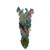
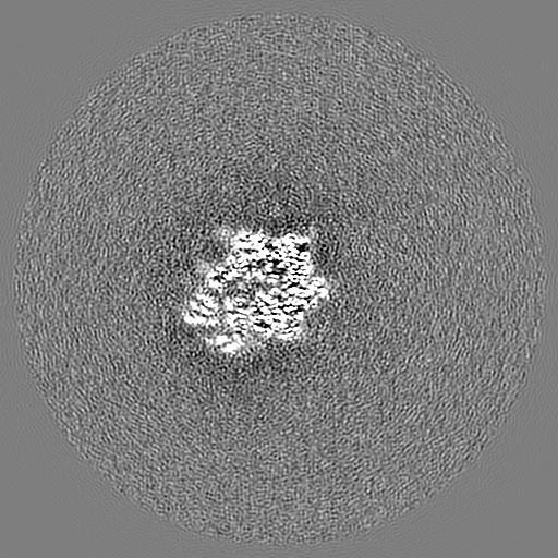
| 画像 | 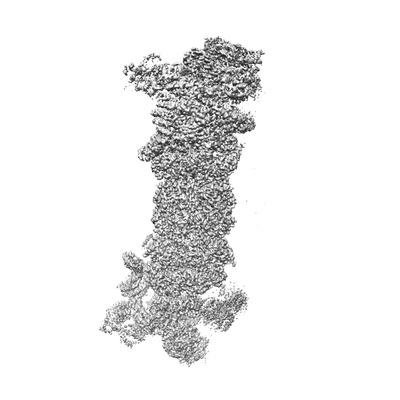 emd_9512.png emd_9512.png | 32.5 KB | ||
| Filedesc metadata | emd-9512.cif.gz | 13.9 KB | ||
| アーカイブディレクトリ |  http://ftp.pdbj.org/pub/emdb/structures/EMD-9512ftp://ftp.pdbj.org/pub/emdb/structures/EMD-9512 http://ftp.pdbj.org/pub/emdb/structures/EMD-9512ftp://ftp.pdbj.org/pub/emdb/structures/EMD-9512 | HTTPS FTP |
-関連構造データ


-リンク
| EMDBのページ | EMDB (EBI/PDBe) / EMDataResource |
|---|---|
| 「今月の分子」の関連する項目 |


-マップ
| ファイル | ダウンロード / ファイル: emd_9512.map.gz / 形式: CCP4 / 大きさ: 512 MB / タイプ: IMAGE STORED AS FLOATING POINT NUMBER (4 BYTES) | ||||||||||||||||||||||||||||||||||||||||||||||||||||||||||||||||||||
|---|---|---|---|---|---|---|---|---|---|---|---|---|---|---|---|---|---|---|---|---|---|---|---|---|---|---|---|---|---|---|---|---|---|---|---|---|---|---|---|---|---|---|---|---|---|---|---|---|---|---|---|---|---|---|---|---|---|---|---|---|---|---|---|---|---|---|---|---|---|
| 注釈 | with C2 symmetry | ||||||||||||||||||||||||||||||||||||||||||||||||||||||||||||||||||||
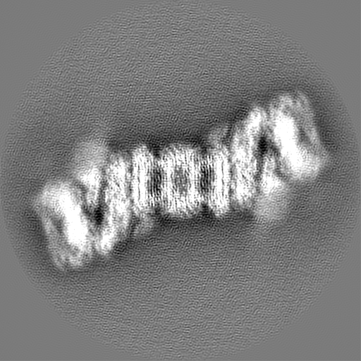
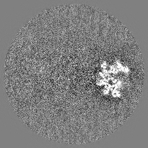
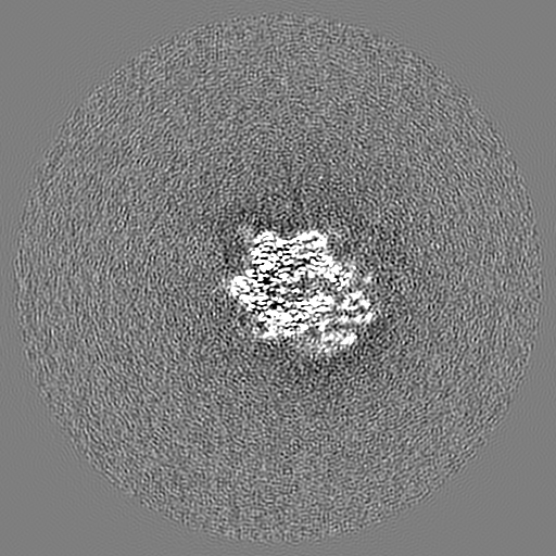
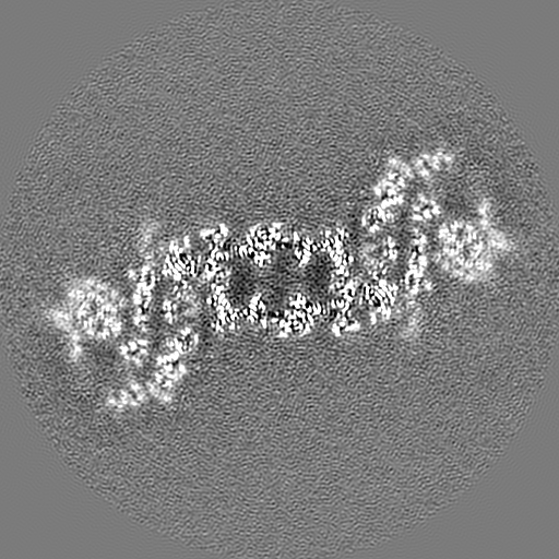
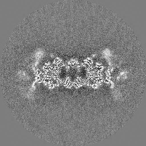
| 投影像・断面図 | 画像のコントロール
画像は Spider により作成 | ||||||||||||||||||||||||||||||||||||||||||||||||||||||||||||||||||||
| ボクセルのサイズ | X=Y=Z: 1.07 Å | ||||||||||||||||||||||||||||||||||||||||||||||||||||||||||||||||||||
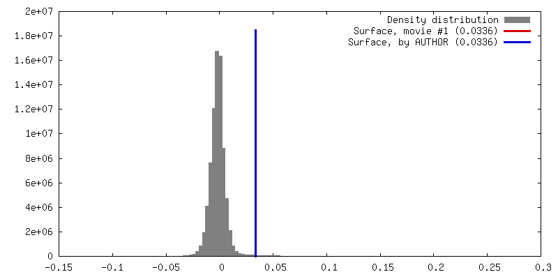
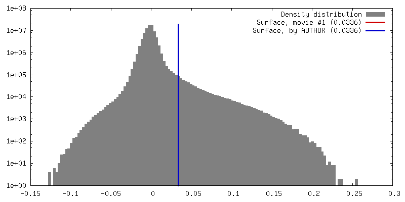
| 密度 |
| ||||||||||||||||||||||||||||||||||||||||||||||||||||||||||||||||||||
| 対称性 | 空間群: 1 | ||||||||||||||||||||||||||||||||||||||||||||||||||||||||||||||||||||
| 詳細 | EMDB XML:
CCP4マップ ヘッダ情報:
| ||||||||||||||||||||||||||||||||||||||||||||||||||||||||||||||||||||
 Z (Sec.)
 Y (Row.)
 X (Col.)


-添付データ
- 試料の構成要素
+全体 : human 26S proteasome
+超分子 #1: human 26S proteasome
+分子 #1: 26S protease regulatory subunit 4
+分子 #2: 26S protease regulatory subunit 7
+分子 #3: 26S protease regulatory subunit 10B
+分子 #4: 26S protease regulatory subunit 6A
+分子 #5: 26S protease regulatory subunit 8
+分子 #6: 26S protease regulatory subunit 6B
+分子 #7: 26S proteasome non-ATPase regulatory subunit 1
+分子 #8: 26S proteasome non-ATPase regulatory subunit 13
+分子 #9: 26S proteasome non-ATPase regulatory subunit 12
+分子 #10: 26S proteasome non-ATPase regulatory subunit 11
+分子 #11: 26S proteasome non-ATPase regulatory subunit 6
+分子 #12: 26S proteasome non-ATPase regulatory subunit 3
+分子 #13: 26S proteasome non-ATPase regulatory subunit 8
+分子 #14: 26S proteasome non-ATPase regulatory subunit 7
+分子 #15: 26S proteasome non-ATPase regulatory subunit 14
+分子 #16: 26S proteasome non-ATPase regulatory subunit 4
+分子 #17: 26S proteasome complex subunit DSS1
+分子 #18: 26S proteasome non-ATPase regulatory subunit 2
+分子 #19: Proteasome subunit alpha type-6
+分子 #20: Proteasome subunit alpha type-2
+分子 #21: Proteasome subunit alpha type-4
+分子 #22: Proteasome subunit alpha type-7
+分子 #23: Proteasome subunit alpha type-5
+分子 #24: Proteasome subunit alpha type-1
+分子 #25: Proteasome subunit alpha type-3
+分子 #26: Proteasome subunit beta type-6
+分子 #27: Proteasome subunit beta type-7
+分子 #28: Proteasome subunit beta type-3
+分子 #29: Proteasome subunit beta type-2
+分子 #30: Proteasome subunit beta type-5
+分子 #31: Proteasome subunit beta type-1
+分子 #32: Proteasome subunit beta type-4
+分子 #33: ADENOSINE-5'-DIPHOSPHATE

-実験情報
-構造解析
| 手法 | クライオ電子顕微鏡法 |
|---|---|
 解析 解析 | 単粒子再構成法 |
| 試料の集合状態 | particle |
-試料調製
| 濃度 | 1 mg/mL | ||||||||||||
|---|---|---|---|---|---|---|---|---|---|---|---|---|---|
| 緩衝液 | pH: 8 構成要素:
| ||||||||||||
| グリッド | 材質: COPPER / 支持フィルム - 材質: CARBON / 支持フィルム - トポロジー: CONTINUOUS / 支持フィルム - Film thickness: 3 / 前処理 - タイプ: GLOW DISCHARGE / 前処理 - 時間: 30 sec. / 前処理 - 雰囲気: AIR | ||||||||||||
| 凍結 | 凍結剤: ETHANE / チャンバー内湿度: 100 % / チャンバー内温度: 281 K / 装置: FEI VITROBOT MARK IV / 詳細: blot for 2 seconds before plunging. | ||||||||||||
| 詳細 | This sample was monodisperse. |
- 電子顕微鏡法
| 顕微鏡 | FEI TITAN KRIOS |
|---|---|
| 温度 | 最低: 70.0 K |
| 詳細 | Preliminary grid screening was performed manually |
| 撮影 | フィルム・検出器のモデル: FEI FALCON II (4k x 4k) デジタル化 - サイズ - 横: 4096 pixel / デジタル化 - サイズ - 縦: 4096 pixel / デジタル化 - 画像ごとのフレーム数: 1-26 / 平均露光時間: 1.6 sec. / 平均電子線量: 37.0 e/Å2 |
| 電子線 | 加速電圧: 300 kV / 電子線源:  FIELD EMISSION GUN FIELD EMISSION GUN |
| 電子光学系 | 最大 デフォーカス(補正後): 0.0026000000000000003 µm 最小 デフォーカス(補正後): 0.0016 µm / 照射モード: FLOOD BEAM / 撮影モード: BRIGHT FIELD / Cs: 2.7 mm |
| 試料ステージ | 試料ホルダーモデル: FEI TITAN KRIOS AUTOGRID HOLDER ホルダー冷却材: NITROGEN |
| 実験機器 |  モデル: Titan Krios / 画像提供: FEI Company |