 Movie
Movie Controller


+ Open data

- Basic information
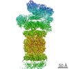
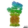
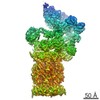
| Entry | Database: EMDB / ID: EMD-9511 | ||||||||||||
|---|---|---|---|---|---|---|---|---|---|---|---|---|---|
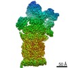
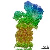
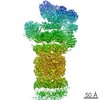
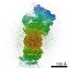
| Title | Cryo-EM map of the human 26S proteasome bound to USP14_UbAl | ||||||||||||
 Map data Map data | Human 26S proteasome bound to USP14-UbAl | ||||||||||||
 Sample Sample |
| ||||||||||||
 Keywords Keywords | protein complex / human proteasome / HYDROLASE | ||||||||||||
| Function / homology |  Function and homology information Function and homology informationnegative regulation of ERAD pathway / regulation of chemotaxis / deubiquitinase activity / protein K48-linked deubiquitination / thyrotropin-releasing hormone receptor binding / nuclear proteasome complex / host-mediated perturbation of viral transcription / positive regulation of inclusion body assembly / Impaired BRCA2 translocation to the nucleus / Impaired BRCA2 binding to SEM1 (DSS1) ...negative regulation of ERAD pathway / regulation of chemotaxis / deubiquitinase activity / protein K48-linked deubiquitination / thyrotropin-releasing hormone receptor binding / nuclear proteasome complex / host-mediated perturbation of viral transcription / positive regulation of inclusion body assembly / Impaired BRCA2 translocation to the nucleus / Impaired BRCA2 binding to SEM1 (DSS1) / meiosis I / proteasome accessory complex / purine ribonucleoside triphosphate binding / integrator complex / proteasome regulatory particle / cytosolic proteasome complex / positive regulation of proteasomal protein catabolic process / hypothalamus gonadotrophin-releasing hormone neuron development / proteasome-activating activity / Antigen processing: Ub, ATP-independent proteasomal degradation / female meiosis I / proteasome regulatory particle, lid subcomplex / proteasome regulatory particle, base subcomplex / positive regulation of protein monoubiquitination / fat pad development / mitochondrion transport along microtubule / sperm glycocalyx / protein K63-linked deubiquitination / negative regulation of programmed cell death / metal-dependent deubiquitinase activity / Regulation of ornithine decarboxylase (ODC) / perinuclear theca / proteasome core complex / Proteasome assembly / endopeptidase inhibitor activity / seminiferous tubule development / Cross-presentation of soluble exogenous antigens (endosomes) / transcription factor binding / K63-linked deubiquitinase activity / Somitogenesis / Homologous DNA Pairing and Strand Exchange / Defective homologous recombination repair (HRR) due to BRCA1 loss of function / Defective HDR through Homologous Recombination Repair (HRR) due to PALB2 loss of BRCA1 binding function / Defective HDR through Homologous Recombination Repair (HRR) due to PALB2 loss of BRCA2/RAD51/RAD51C binding function / Resolution of D-loop Structures through Synthesis-Dependent Strand Annealing (SDSA) / female gonad development / Resolution of D-loop Structures through Holliday Junction Intermediates / proteasome binding / Impaired BRCA2 binding to RAD51 / regulation of protein catabolic process / myofibril / male meiosis I / proteasome storage granule / proteasomal ubiquitin-independent protein catabolic process / sperm head-tail coupling apparatus / positive regulation of RNA polymerase II transcription preinitiation complex assembly / general transcription initiation factor binding / Presynaptic phase of homologous DNA pairing and strand exchange / positive regulation of intrinsic apoptotic signaling pathway by p53 class mediator / blastocyst development / protein deubiquitination / immune system process / polyubiquitin modification-dependent protein binding / proteasome endopeptidase complex / NF-kappaB binding / proteasome core complex, beta-subunit complex / negative regulation of ubiquitin-dependent protein catabolic process / endopeptidase activator activity / threonine-type endopeptidase activity / proteasome core complex, alpha-subunit complex / mRNA export from nucleus / proteasome assembly / SARS-CoV-1 targets host intracellular signalling and regulatory pathways / presynaptic cytosol / enzyme regulator activity / energy homeostasis / neuron projection morphogenesis / ERAD pathway / regulation of proteasomal protein catabolic process / inclusion body / Maturation of protein E / Maturation of protein E / ER Quality Control Compartment (ERQC) / Myoclonic epilepsy of Lafora / FLT3 signaling by CBL mutants / IRAK2 mediated activation of TAK1 complex / Alpha-protein kinase 1 signaling pathway / Glycogen synthesis / IRAK1 recruits IKK complex / IRAK1 recruits IKK complex upon TLR7/8 or 9 stimulation / Prevention of phagosomal-lysosomal fusion / Endosomal Sorting Complex Required For Transport (ESCRT) / Membrane binding and targetting of GAG proteins / Regulation of TBK1, IKKε (IKBKE)-mediated activation of IRF3, IRF7 / Negative regulation of FLT3 / PTK6 Regulates RTKs and Their Effectors AKT1 and DOK1 / Regulation of TBK1, IKKε-mediated activation of IRF3, IRF7 upon TLR3 ligation / IRAK2 mediated activation of TAK1 complex upon TLR7/8 or 9 stimulation / Constitutive Signaling by NOTCH1 HD Domain Mutants / NOTCH2 Activation and Transmission of Signal to the Nucleus Similarity search - Function | ||||||||||||
| Biological species |  Homo sapiens (human) Homo sapiens (human) | ||||||||||||
| Method | single particle reconstruction / cryo EM / Resolution: 4.35 Å | ||||||||||||
 Authors Authors | Huang XL / Luan B / Wu JP / Shi YG | ||||||||||||
| Funding support |  China, 3 items China, 3 items
| ||||||||||||
 Citation Citation | Journal: Nat Struct Mol Biol / Year: 2016 Title: An atomic structure of the human 26S proteasome. Authors: Xiuliang Huang / Bai Luan / Jianping Wu / Yigong Shi / Abstract: We report the cryo-EM structure of the human 26S proteasome at an average resolution of 3.5 Å, allowing atomic modeling of 28 subunits in the core particle (CP) and 18 subunits in the regulatory ...We report the cryo-EM structure of the human 26S proteasome at an average resolution of 3.5 Å, allowing atomic modeling of 28 subunits in the core particle (CP) and 18 subunits in the regulatory particle (RP). The C-terminal residues of Rpt3 and Rpt5 subunits in the RP can be seen inserted into surface pockets formed between adjacent α subunits in the CP. Each of the six Rpt subunits contains a bound nucleotide, and the central gate of the CP α-ring is closed despite RP association. The six pore 1 loops in the Rpt ring are arranged similarly to a spiral staircase along the axial channel of substrate transport, which is constricted by the pore 2 loops. We also determined the cryo-EM structure of the human proteasome bound to the deubiquitinating enzyme USP14 at 4.35-Å resolution. Together, our structures provide a framework for mechanistic understanding of eukaryotic proteasome function. | ||||||||||||
| History |
|
- Structure visualization
| Movie |
Movie viewer |
|---|---|
| Structure viewer | EM map: SurfViewMolmilJmol/JSmol |
| Supplemental images |

- Downloads & links
-EMDB archive
| Map data | emd_9511.map.gz | 480 MB | EMDB map data format | |
|---|---|---|---|---|
| Header (meta data) | emd-9511-v30.xmlemd-9511.xml | 60.1 KB 60.1 KB | Display Display | EMDB header |
| Images | 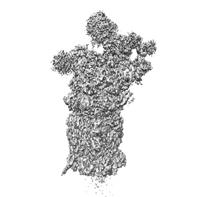 emd_9511.png emd_9511.png | 45.1 KB | ||
| Filedesc metadata | emd-9511.cif.gz | 16 KB | ||
| Archive directory |  http://ftp.pdbj.org/pub/emdb/structures/EMD-9511ftp://ftp.pdbj.org/pub/emdb/structures/EMD-9511 http://ftp.pdbj.org/pub/emdb/structures/EMD-9511ftp://ftp.pdbj.org/pub/emdb/structures/EMD-9511 | HTTPS FTP |
-Related structure data
| Related structure data |  5gjqMC  9507C 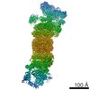 9508C 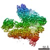 9509C  9510C 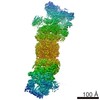 9512C 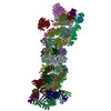 5gjrC C: citing same article ( M: atomic model generated by this map |
|---|---|
| Similar structure data |
-Links
| EMDB pages | EMDB (EBI/PDBe) / EMDataResource |
|---|---|
| Related items in Molecule of the Month |


-Map
| File | Download / File: emd_9511.map.gz / Format: CCP4 / Size: 512 MB / Type: IMAGE STORED AS FLOATING POINT NUMBER (4 BYTES) | ||||||||||||||||||||||||||||||||||||||||||||||||||||||||||||||||||||
|---|---|---|---|---|---|---|---|---|---|---|---|---|---|---|---|---|---|---|---|---|---|---|---|---|---|---|---|---|---|---|---|---|---|---|---|---|---|---|---|---|---|---|---|---|---|---|---|---|---|---|---|---|---|---|---|---|---|---|---|---|---|---|---|---|---|---|---|---|---|
| Annotation | Human 26S proteasome bound to USP14-UbAl | ||||||||||||||||||||||||||||||||||||||||||||||||||||||||||||||||||||
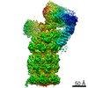
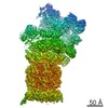
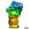
| Projections & slices | Image control
Images are generated by Spider. | ||||||||||||||||||||||||||||||||||||||||||||||||||||||||||||||||||||
| Voxel size | X=Y=Z: 1.07 Å | ||||||||||||||||||||||||||||||||||||||||||||||||||||||||||||||||||||
| Density |
| ||||||||||||||||||||||||||||||||||||||||||||||||||||||||||||||||||||
| Symmetry | Space group: 1 | ||||||||||||||||||||||||||||||||||||||||||||||||||||||||||||||||||||
| Details | EMDB XML:
CCP4 map header:
| ||||||||||||||||||||||||||||||||||||||||||||||||||||||||||||||||||||
 Z (Sec.)
 Y (Row.)
 X (Col.)


-Supplemental data
- Sample components
+Entire : human 26S proteasome bound to USP14-UbAl
+Supramolecule #1: human 26S proteasome bound to USP14-UbAl
+Macromolecule #1: Proteasome subunit beta type-6
+Macromolecule #2: Proteasome subunit alpha type-6
+Macromolecule #3: Proteasome subunit beta type-7
+Macromolecule #4: Proteasome subunit alpha type-2
+Macromolecule #5: Proteasome subunit beta type-3
+Macromolecule #6: Proteasome subunit alpha type-4
+Macromolecule #7: Proteasome subunit beta type-2
+Macromolecule #8: Proteasome subunit alpha type-7
+Macromolecule #9: Proteasome subunit beta type-5
+Macromolecule #10: Proteasome subunit alpha type-5
+Macromolecule #11: Proteasome subunit beta type-1
+Macromolecule #12: Proteasome subunit alpha type-1
+Macromolecule #13: Proteasome subunit beta type-4
+Macromolecule #14: 26S protease regulatory subunit 7
+Macromolecule #15: 26S protease regulatory subunit 4
+Macromolecule #16: 26S protease regulatory subunit 8
+Macromolecule #17: 26S protease regulatory subunit 6B
+Macromolecule #18: 26S protease regulatory subunit 10B
+Macromolecule #19: 26S protease regulatory subunit 6A
+Macromolecule #20: 26S proteasome non-ATPase regulatory subunit 1
+Macromolecule #21: Proteasome subunit alpha type-3
+Macromolecule #22: 26S proteasome non-ATPase regulatory subunit 13
+Macromolecule #23: 26S proteasome non-ATPase regulatory subunit 12
+Macromolecule #24: 26S proteasome non-ATPase regulatory subunit 11
+Macromolecule #25: 26S proteasome non-ATPase regulatory subunit 6
+Macromolecule #26: 26S proteasome non-ATPase regulatory subunit 3
+Macromolecule #27: 26S proteasome non-ATPase regulatory subunit 8
+Macromolecule #28: 26S proteasome non-ATPase regulatory subunit 7
+Macromolecule #29: 26S proteasome non-ATPase regulatory subunit 14
+Macromolecule #30: 26S proteasome non-ATPase regulatory subunit 4
+Macromolecule #31: 26S proteasome complex subunit DSS1
+Macromolecule #32: 26S proteasome non-ATPase regulatory subunit 2
+Macromolecule #33: Ubiquitin carboxyl-terminal hydrolase 14

+Macromolecule #34: Polyubiquitin-B
+Macromolecule #35: ADENOSINE-5'-DIPHOSPHATE

-Experimental details
-Structure determination
| Method | cryo EM |
|---|---|
 Processing Processing | single particle reconstruction |
| Aggregation state | particle |
-Sample preparation
| Concentration | 1 mg/mL | ||||||||||||
|---|---|---|---|---|---|---|---|---|---|---|---|---|---|
| Buffer | pH: 8 Component:
| ||||||||||||
| Grid | Material: COPPER / Support film - Material: CARBON / Support film - topology: CONTINUOUS / Support film - Film thickness: 3 / Pretreatment - Type: GLOW DISCHARGE / Pretreatment - Time: 30 sec. / Pretreatment - Atmosphere: AIR | ||||||||||||
| Vitrification | Cryogen name: ETHANE / Chamber humidity: 100 % / Chamber temperature: 281 K / Instrument: FEI VITROBOT MARK IV / Details: blot for 2 seconds before plunging. | ||||||||||||
| Details | This sample was monodisperse. |
- Electron microscopy
| Microscope | FEI TITAN KRIOS |
|---|---|
| Temperature | Min: 70.0 K |
| Details | Preliminary grid screening was performed manually |
| Image recording | Film or detector model: FEI FALCON II (4k x 4k) / Digitization - Dimensions - Width: 4096 pixel / Digitization - Dimensions - Height: 4096 pixel / Digitization - Frames/image: 1-26 / Average exposure time: 1.6 sec. / Average electron dose: 37.0 e/Å2 |
| Electron beam | Acceleration voltage: 300 kV / Electron source:  FIELD EMISSION GUN FIELD EMISSION GUN |
| Electron optics | Illumination mode: FLOOD BEAM / Imaging mode: BRIGHT FIELD / Cs: 2.7 mm |
| Sample stage | Specimen holder model: FEI TITAN KRIOS AUTOGRID HOLDER / Cooling holder cryogen: NITROGEN |
| Experimental equipment |  Model: Titan Krios / Image courtesy: FEI Company |
