- EMDB-7831: Cryo-EM structure of FLNaABD E254K bound to phalloidin-stabilized... -
+
Open data
ID or keywords:
Loading...
-
Basic information
Entry
Database: EMDB / ID: EMD-7831
Title
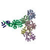
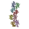
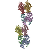
Cryo-EM structure of FLNaABD E254K bound to phalloidin-stabilized F-actin
Map data
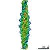
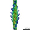
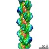
Symmetrized map of FLNaABD-E254K bound to phalloidin-stabilized F-actin. This map has been low-pass filtered to 3.6 A and sharpened with a B-factor of -150.
Sample
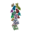
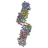
Complex: Helical complex of FLNaABD E254K bound to phalloidin-stabilized F-actin
Protein or peptide: Filamin-A
Protein or peptide: Actin, alpha skeletal muscle
Protein or peptide: Phalloidin
Ligand: ADENOSINE-5'-DIPHOSPHATE
Ligand: MAGNESIUM ION
Keywords
Actin-binding domain / Actin crosslinker / STRUCTURAL PROTEIN
Function / homology
Function and homology information
regulation of membrane repolarization during atrial cardiac muscle cell action potential / regulation of membrane repolarization during cardiac muscle cell action potential / establishment of Sertoli cell barrier / Myb complex / glycoprotein Ib-IX-V complex / adenylate cyclase-inhibiting dopamine receptor signaling pathway / formation of radial glial scaffolds / positive regulation of integrin-mediated signaling pathway / Regulation of CDH1 Function / blood coagulation, intrinsic pathway ...regulation of membrane repolarization during atrial cardiac muscle cell action potential / regulation of membrane repolarization during cardiac muscle cell action potential / establishment of Sertoli cell barrier / Myb complex / glycoprotein Ib-IX-V complex / adenylate cyclase-inhibiting dopamine receptor signaling pathway / formation of radial glial scaffolds / positive regulation of integrin-mediated signaling pathway / Regulation of CDH1 Function / blood coagulation, intrinsic pathway / tubulin deacetylation / OAS antiviral response / actin crosslink formation / Striated Muscle Contraction / positive regulation of actin filament bundle assembly / positive regulation of neuron migration / protein localization to bicellular tight junction / Cell-extracellular matrix interactions / positive regulation of potassium ion transmembrane transport / positive regulation of platelet activation / apical dendrite / Fc-gamma receptor I complex binding / positive regulation of neural precursor cell proliferation / podosome / protein localization to cell surface / wound healing, spreading of cells / negative regulation of transcription by RNA polymerase I / megakaryocyte development / GP1b-IX-V activation signalling / SMAD binding / receptor clustering / cortical cytoskeleton / semaphorin-plexin signaling pathway / RHO GTPases activate PAKs / striated muscle thin filament / skeletal muscle thin filament assembly / cilium assembly / : / mitotic spindle assembly / potassium channel regulator activity / skeletal muscle fiber development / stress fiber / release of sequestered calcium ion into cytosol / positive regulation of substrate adhesion-dependent cell spreading / regulation of cell migration / dendritic shaft / protein localization to plasma membrane / actin filament / establishment of protein localization / protein sequestering activity / negative regulation of protein catabolic process / cerebral cortex development / positive regulation of protein import into nucleus / G protein-coupled receptor binding / mRNA transcription by RNA polymerase II / platelet aggregation / Hydrolases; Acting on acid anhydrides; Acting on acid anhydrides to facilitate cellular and subcellular movement / Z disc / small GTPase binding / kinase binding / cell-cell junction / actin filament binding / Platelet degranulation / actin cytoskeleton / toxin activity / growth cone / actin cytoskeleton organization / GTPase binding / DNA-binding transcription factor binding / perikaryon / transmembrane transporter binding / positive regulation of canonical NF-kappaB signal transduction / postsynapse / protein stabilization / cadherin binding / focal adhesion / hydrolase activity / negative regulation of apoptotic process / nucleolus / perinuclear region of cytoplasm / glutamatergic synapse / protein homodimerization activity / RNA binding / extracellular exosome / extracellular region / ATP binding / membrane / nucleus / plasma membrane / cytoplasm / cytosol Similarity search - Function
Journal: Nat Struct Mol Biol / Year: 2018 Title: Structural basis of the filamin A actin-binding domain interaction with F-actin. Authors: Daniel V Iwamoto / Andrew Huehn / Bertrand Simon / Clotilde Huet-Calderwood / Massimiliano Baldassarre / Charles V Sindelar / David A Calderwood / Abstract: Actin-cross-linking proteins assemble actin filaments into higher-order structures essential for orchestrating cell shape, adhesion, and motility. Missense mutations in the tandem calponin homology ...Actin-cross-linking proteins assemble actin filaments into higher-order structures essential for orchestrating cell shape, adhesion, and motility. Missense mutations in the tandem calponin homology domains of their actin-binding domains (ABDs) underlie numerous genetic diseases, but a molecular understanding of these pathologies is hampered by the lack of high-resolution structures of any actin-cross-linking protein bound to F-actin. Here, taking advantage of a high-affinity, disease-associated mutant of the human filamin A (FLNa) ABD, we combine cryo-electron microscopy and functional studies to reveal at near-atomic resolution how the first calponin homology domain (CH1) and residues immediately N-terminal to it engage actin. We further show that reorientation of CH2 relative to CH1 is required to avoid clashes with actin and to expose F-actin-binding residues on CH1. Our data explain localization of disease-associated loss-of-function mutations to FLNaCH1 and gain-of-function mutations to the regulatory FLNaCH2. Sequence conservation argues that this provides a general model for ABD-F-actin binding.
History
Deposition
Apr 26, 2018
-
Header (metadata) release
Aug 8, 2018
-
Map release
Sep 19, 2018
-
Update
Nov 15, 2023
-
Current status
Nov 15, 2023
Processing site: RCSB / Status: Released
-
Structure visualization
Movie
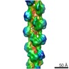
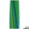
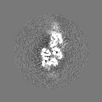
Surface view with section colored by density value
Download / File: emd_7831.map.gz / Format: CCP4 / Size: 35.3 MB / Type: IMAGE STORED AS FLOATING POINT NUMBER (4 BYTES)
Annotation
Symmetrized map of FLNaABD-E254K bound to phalloidin-stabilized F-actin. This map has been low-pass filtered to 3.6 A and sharpened with a B-factor of -150.
Symmetrized map of FLNaABD-E254K bound to phalloidin-stabilized F-actin. Low-pass filtered to 10 A and sharpened with a B-factor of -150. Masked to display the FLNaABD CH2 domain cryo-EM density.
Independently refined half map(2) of FLNaABD-E254K bound to phalloidin-stabilized F-actin. This half map was symmetrized using parameters calculated from the full map.
Independently refined half map(1) of FLNaABD-E254K bound to phalloidin-stabilized F-actin. This half map was symmetrized using parameters calculated from the full map.
Film or detector model: GATAN K2 SUMMIT (4k x 4k) / Detector mode: SUPER-RESOLUTION / Digitization - Frames/image: 1-40 / Number grids imaged: 2 / Number real images: 2140 / Average exposure time: 12.0 sec. / Average electron dose: 50.0 e/Å2 Details: Micrographs from only one of the grids were used in the reconstruction. From that grid, only micrographs where Gctf detected signal at resolutions better than 4A were used in the reconstruction (~15%).
Electron beam
Acceleration voltage: 300 kV / Electron source: FIELD EMISSION GUN
In the structure databanks used in Yorodumi, some data are registered as the other names, "COVID-19 virus" and "2019-nCoV". Here are the details of the virus and the list of structure data.
Jan 31, 2019. EMDB accession codes are about to change! (news from PDBe EMDB page)
EMDB accession codes are about to change! (news from PDBe EMDB page)
The allocation of 4 digits for EMDB accession codes will soon come to an end. Whilst these codes will remain in use, new EMDB accession codes will include an additional digit and will expand incrementally as the available range of codes is exhausted. The current 4-digit format prefixed with “EMD-” (i.e. EMD-XXXX) will advance to a 5-digit format (i.e. EMD-XXXXX), and so on. It is currently estimated that the 4-digit codes will be depleted around Spring 2019, at which point the 5-digit format will come into force.
The EM Navigator/Yorodumi systems omit the EMD- prefix.
Related info.:Q: What is EMD? / ID/Accession-code notation in Yorodumi/EM Navigator
Yorodumi is a browser for structure data from EMDB, PDB, SASBDB, etc.
This page is also the successor to EM Navigator detail page, and also detail information page/front-end page for Omokage search.
The word "yorodu" (or yorozu) is an old Japanese word meaning "ten thousand". "mi" (miru) is to see.
Related info.:EMDB / PDB / SASBDB / Comparison of 3 databanks / Yorodumi Search / Aug 31, 2016. New EM Navigator & Yorodumi / Yorodumi Papers / Jmol/JSmol / Function and homology information / Changes in new EM Navigator and Yorodumi
 Movie
Movie Controller

 Yorodumi
 Open data
 Basic information
 Map data
 Sample
 Keywords
 Function and homology information
 Homo sapiens (human) /
 Amanita phalloides (death cap)
 Authors
 Citation

 Structure visualization
 Downloads & links
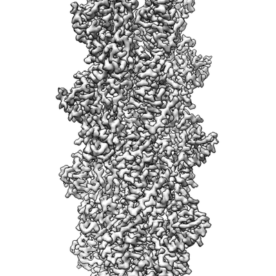 emd_7831.png
 http://ftp.pdbj.org/pub/emdb/structures/EMD-7831


 Z (Sec.)
 Y (Row.)
 X (Col.)


 Sample components

 Processing
 Electron microscopy
 FIELD EMISSION GUN