- EMDB-6988: cryo-em structure of alpha-synuclein fiber -
+
Open data
ID or keywords:
Loading...
-
Basic information
Entry
Database: EMDB / ID: EMD-6988
Title
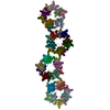
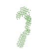
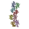
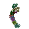
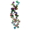
cryo-em structure of alpha-synuclein fiber
Map data
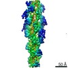
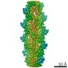
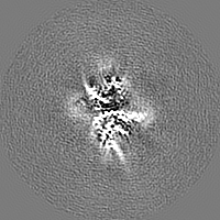
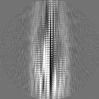
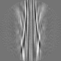
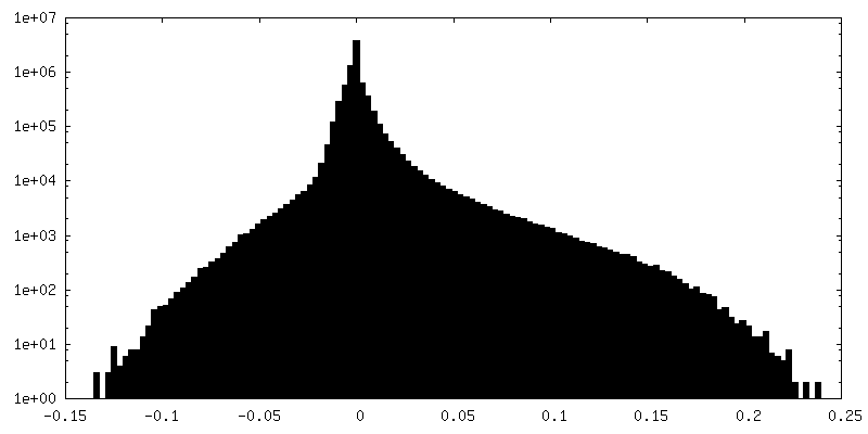
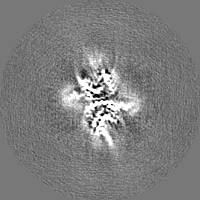
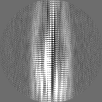
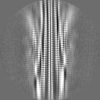
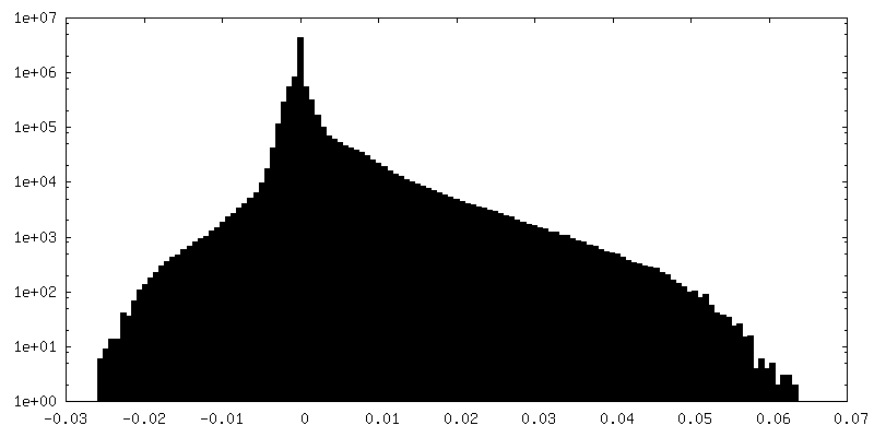
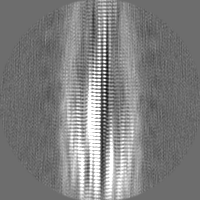
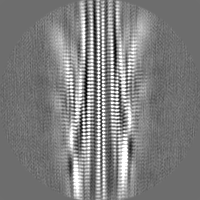
Density map calculated by applying a helical symmetry to the segments in the middle of 3D reconstruction. The twist angle is 179.64 degree and the rise is 3.39 angstrom.
Sample
Complex: alpha-synuclein fiber
Protein or peptide: Alpha-synuclein
Keywords
alpha-syn fiber / Parkinson disease / PROTEIN FIBRIL
Function / homology
Function and homology information
negative regulation of mitochondrial electron transport, NADH to ubiquinone / negative regulation of dopamine uptake involved in synaptic transmission / negative regulation of norepinephrine uptake / response to desipramine / positive regulation of SNARE complex assembly / positive regulation of hydrogen peroxide catabolic process / supramolecular fiber / regulation of synaptic vesicle recycling / negative regulation of chaperone-mediated autophagy / regulation of reactive oxygen species biosynthetic process ...negative regulation of mitochondrial electron transport, NADH to ubiquinone / negative regulation of dopamine uptake involved in synaptic transmission / negative regulation of norepinephrine uptake / response to desipramine / positive regulation of SNARE complex assembly / positive regulation of hydrogen peroxide catabolic process / supramolecular fiber / regulation of synaptic vesicle recycling / negative regulation of chaperone-mediated autophagy / regulation of reactive oxygen species biosynthetic process / positive regulation of protein localization to cell periphery / negative regulation of exocytosis / dopamine biosynthetic process / dopamine uptake involved in synaptic transmission / response to iron(II) ion / negative regulation of dopamine metabolic process / negative regulation of platelet-derived growth factor receptor signaling pathway / SNARE complex assembly / negative regulation of thrombin-activated receptor signaling pathway / Lewy body / negative regulation of microtubule polymerization / synaptic vesicle priming / regulation of norepinephrine uptake / transporter regulator activity / protein kinase inhibitor activity / positive regulation of inositol phosphate biosynthetic process / synaptic vesicle transport / regulation of dopamine secretion / positive regulation of receptor recycling / cuprous ion binding / positive regulation of exocytosis / nuclear outer membrane / dynein complex binding / synaptic transmission, dopaminergic / synaptic vesicle exocytosis / response to magnesium ion / positive regulation of endocytosis / negative regulation of serotonin uptake / kinesin binding / cysteine-type endopeptidase inhibitor activity / regulation of presynapse assembly / synaptic vesicle endocytosis / alpha-tubulin binding / beta-tubulin binding / phospholipase binding / behavioral response to cocaine / supramolecular fiber organization / cellular response to fibroblast growth factor stimulus / response to type II interferon / cellular response to epinephrine stimulus / inclusion body / Hsp70 protein binding / response to interleukin-1 / axon terminus / cellular response to copper ion / positive regulation of release of sequestered calcium ion into cytosol / enzyme inhibitor activity / regulation of microtubule cytoskeleton organization / SNARE binding / glutathione metabolic process / protein tetramerization / protein sequestering activity / phosphoprotein binding / receptor internalization / tubulin binding / microglial cell activation / ferrous iron binding / phospholipid binding / PKR-mediated signaling / synapse organization / protein destabilization / tau protein binding / enzyme activator activity / terminal bouton / positive regulation of inflammatory response / actin cytoskeleton / synaptic vesicle membrane / growth cone / actin binding / cellular response to oxidative stress / response to lipopolysaccharide / histone binding / cell cortex / microtubule binding / amyloid fibril formation / negative regulation of neuron apoptotic process / mitochondrial outer membrane / lysosome / oxidoreductase activity / mitochondrial inner membrane / transcription cis-regulatory region binding / positive regulation of apoptotic process / ribosome / mitochondrial matrix / Amyloid fiber formation / copper ion binding / protein domain specific binding / axon / neuronal cell body / calcium ion binding Similarity search - Function
Journal: Cell Res / Year: 2018 Title: Amyloid fibril structure of α-synuclein determined by cryo-electron microscopy. Authors: Yaowang Li / Chunyu Zhao / Feng Luo / Zhenying Liu / Xinrui Gui / Zhipu Luo / Xiang Zhang / Dan Li / Cong Liu / Xueming Li / Abstract: α-Synuclein (α-syn) amyloid fibrils are the major component of Lewy bodies, which are the pathological hallmark of Parkinson's disease (PD) and other synucleinopathies. High-resolution structure of ...α-Synuclein (α-syn) amyloid fibrils are the major component of Lewy bodies, which are the pathological hallmark of Parkinson's disease (PD) and other synucleinopathies. High-resolution structure of α-syn fibril is important for understanding its assembly and pathological mechanism. Here, we determined a fibril structure of full-length α-syn (1-140) at the resolution of 3.07 Å by cryo-electron microscopy (cryo-EM). The fibrils are cytotoxic, and transmissible to induce endogenous α-syn aggregation in primary neurons. Based on the reconstructed cryo-EM density map, we were able to unambiguously build the fibril structure comprising residues 37-99. The α-syn amyloid fibril structure shows two protofilaments intertwining along an approximate 2 screw axis into a left-handed helix. Each protofilament features a Greek key-like topology. Remarkably, five out of the six early-onset PD familial mutations are located at the dimer interface of the fibril (H50Q, G51D, and A53T/E) or involved in the stabilization of the protofilament (E46K). Furthermore, these PD mutations lead to the formation of fibrils with polymorphic structures distinct from that of the wild-type. Our study provides molecular insight into the fibrillar assembly of α-syn at the atomic level and sheds light on the molecular pathogenesis caused by familial PD mutations of α-syn.
History
Deposition
Jun 27, 2018
-
Header (metadata) release
Jul 11, 2018
-
Map release
Jul 11, 2018
-
Update
Mar 27, 2024
-
Current status
Mar 27, 2024
Processing site: PDBj / Status: Released
-
Structure visualization
Movie
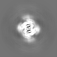
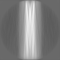
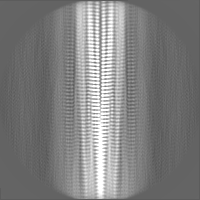
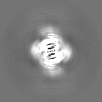
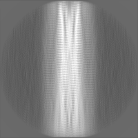
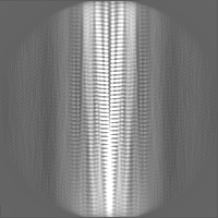
Surface view with section colored by density value
Download / File: emd_6988.map.gz / Format: CCP4 / Size: 30.5 MB / Type: IMAGE STORED AS FLOATING POINT NUMBER (4 BYTES)
Annotation
Density map calculated by applying a helical symmetry to the segments in the middle of 3D reconstruction. The twist angle is 179.64 degree and the rise is 3.39 angstrom.
Model: Quantifoil R1.2/1.3 / Material: COPPER / Mesh: 300 / Pretreatment - Type: GLOW DISCHARGE Details: 4 microl of alpha-syn fibril solution was applied to a glow-discharged holey carbon grid (Quantifoil R1.2/1.3, 300 mesh), blotted for 6 s, and plunge-frozen in liquid ethane using FEI ...Details: 4 microl of alpha-syn fibril solution was applied to a glow-discharged holey carbon grid (Quantifoil R1.2/1.3, 300 mesh), blotted for 6 s, and plunge-frozen in liquid ethane using FEI Vitrobot Mark IV. 95% humidity, 16 degrees
Vitrification
Cryogen name: ETHANE / Chamber humidity: 95 % / Chamber temperature: 289 K / Instrument: FEI VITROBOT MARK IV
-
Electron microscopy
Microscope
FEI TITAN KRIOS
Image recording
Film or detector model: GATAN K2 SUMMIT (4k x 4k) / Detector mode: SUPER-RESOLUTION / Digitization - Frames/image: 1-32 / Average exposure time: 8.0 sec. / Average electron dose: 50.0 e/Å2
Electron beam
Acceleration voltage: 300 kV / Electron source: FIELD EMISSION GUN
Electron optics
Illumination mode: FLOOD BEAM / Imaging mode: BRIGHT FIELD
In the structure databanks used in Yorodumi, some data are registered as the other names, "COVID-19 virus" and "2019-nCoV". Here are the details of the virus and the list of structure data.
Jan 31, 2019. EMDB accession codes are about to change! (news from PDBe EMDB page)
EMDB accession codes are about to change! (news from PDBe EMDB page)
The allocation of 4 digits for EMDB accession codes will soon come to an end. Whilst these codes will remain in use, new EMDB accession codes will include an additional digit and will expand incrementally as the available range of codes is exhausted. The current 4-digit format prefixed with “EMD-” (i.e. EMD-XXXX) will advance to a 5-digit format (i.e. EMD-XXXXX), and so on. It is currently estimated that the 4-digit codes will be depleted around Spring 2019, at which point the 5-digit format will come into force.
The EM Navigator/Yorodumi systems omit the EMD- prefix.
Related info.:Q: What is EMD? / ID/Accession-code notation in Yorodumi/EM Navigator
Yorodumi is a browser for structure data from EMDB, PDB, SASBDB, etc.
This page is also the successor to EM Navigator detail page, and also detail information page/front-end page for Omokage search.
The word "yorodu" (or yorozu) is an old Japanese word meaning "ten thousand". "mi" (miru) is to see.
Related info.:EMDB / PDB / SASBDB / Comparison of 3 databanks / Yorodumi Search / Aug 31, 2016. New EM Navigator & Yorodumi / Yorodumi Papers / Jmol/JSmol / Function and homology information / Changes in new EM Navigator and Yorodumi
 Movie
Movie Controller


 Open data
 Basic information
 Map data
 Sample
 Keywords
 Function and homology information
 Homo sapiens (human)
 Authors
 Citation
 Structure visualization
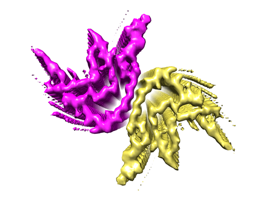
 Downloads & links
 emd_6988.png
 http://ftp.pdbj.org/pub/emdb/structures/EMD-6988
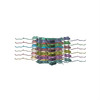


 Z (Sec.)
 Y (Row.)
 X (Col.)


 Sample components
 Processing
 Electron microscopy
 FIELD EMISSION GUN
