 Movie
Movie Controller

[English] 日本語
 Yorodumi
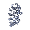
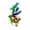
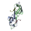
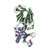
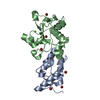
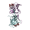
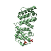
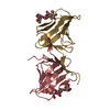
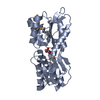
- PDB-3hoc: Structure of the actin-binding domain of human filamin A mutant E254K -
+ Open data

- Basic information
| Entry | Database: PDB / ID: 3hoc | ||||||
|---|---|---|---|---|---|---|---|
| Title | Structure of the actin-binding domain of human filamin A mutant E254K | ||||||
 Components Components | Filamin-A | ||||||
 Keywords Keywords | STRUCTURAL PROTEIN / Calponin homology domain / actin binding domain / Acetylation / Actin-binding / Alternative splicing / Cytoplasm / Cytoskeleton / Disease mutation / Phosphoprotein / Polymorphism | ||||||
| Function / homology |  Function and homology information Function and homology informationregulation of membrane repolarization during atrial cardiac muscle cell action potential / regulation of membrane repolarization during cardiac muscle cell action potential / establishment of Sertoli cell barrier / formation of radial glial scaffolds / Myb complex / adenylate cyclase-inhibiting dopamine receptor signaling pathway / positive regulation of integrin-mediated signaling pathway / blood coagulation, intrinsic pathway / OAS antiviral response / protein localization to bicellular tight junction ...regulation of membrane repolarization during atrial cardiac muscle cell action potential / regulation of membrane repolarization during cardiac muscle cell action potential / establishment of Sertoli cell barrier / formation of radial glial scaffolds / Myb complex / adenylate cyclase-inhibiting dopamine receptor signaling pathway / positive regulation of integrin-mediated signaling pathway / blood coagulation, intrinsic pathway / OAS antiviral response / protein localization to bicellular tight junction / actin crosslink formation / positive regulation of actin filament bundle assembly / positive regulation of neuron migration / tubulin deacetylation / megakaryocyte development / Cell-extracellular matrix interactions / positive regulation of platelet activation / positive regulation of potassium ion transmembrane transport / apical dendrite / Fc-gamma receptor I complex binding / positive regulation of neural precursor cell proliferation / protein localization to cell surface / negative regulation of transcription by RNA polymerase I / podosome / wound healing, spreading of cells / GP1b-IX-V activation signalling / SMAD binding / receptor clustering / cortical cytoskeleton / RHO GTPases activate PAKs / semaphorin-plexin signaling pathway / mitotic spindle assembly / cilium assembly / potassium channel regulator activity / release of sequestered calcium ion into cytosol / positive regulation of substrate adhesion-dependent cell spreading / regulation of cell migration / protein localization to plasma membrane / dendritic shaft / actin filament / establishment of protein localization / negative regulation of protein catabolic process / protein sequestering activity / cerebral cortex development / positive regulation of protein import into nucleus / platelet aggregation / mRNA transcription by RNA polymerase II / G protein-coupled receptor binding / small GTPase binding / kinase binding / Z disc / cell-cell junction / actin filament binding / actin cytoskeleton / Platelet degranulation / growth cone / actin cytoskeleton organization / GTPase binding / DNA-binding transcription factor binding / perikaryon / transmembrane transporter binding / positive regulation of canonical NF-kappaB signal transduction / postsynapse / protein stabilization / cadherin binding / focal adhesion / nucleolus / negative regulation of apoptotic process / perinuclear region of cytoplasm / glutamatergic synapse / protein homodimerization activity / RNA binding / extracellular exosome / extracellular region / membrane / nucleus / plasma membrane / cytosol / cytoplasm Similarity search - Function | ||||||
| Biological species |  Homo sapiens (human) Homo sapiens (human) | ||||||
| Method |  X-RAY DIFFRACTION / MOLECULAR REPLACEMENT / Resolution: 2.3 Å X-RAY DIFFRACTION / MOLECULAR REPLACEMENT / Resolution: 2.3 Å | ||||||
 Authors Authors | Clark, A.R. / Sawyer, G.M. / Robertson, S.P. / Sutherland-Smith, A.J. | ||||||
 Citation Citation | Journal: Hum.Mol.Genet. / Year: 2009 Title: Skeletal dysplasias due to filamin A mutations result from a gain-of-function mechanism distinct from allelic neurological disorders Authors: Clark, A.R. / Sawyer, G.M. / Robertson, S.P. / Sutherland-Smith, A.J. | ||||||
| History |
|
- Structure visualization
| Structure viewer | Molecule: MolmilJmol/JSmol |
|---|
- Downloads & links
-Download
| PDBx/mmCIF format | 3hoc.cif.gz | 186.8 KB | Display | PDBx/mmCIF format |
|---|---|---|---|---|
| PDB format | pdb3hoc.ent.gz | 149.4 KB | Display | PDB format |
| PDBx/mmJSON format | 3hoc.json.gz | Tree view | PDBx/mmJSON format | |
| Others |  Other downloads Other downloads |
-Validation report
| Arichive directory | https://data.pdbj.org/pub/pdb/validation_reports/ho/3hocftp://data.pdbj.org/pub/pdb/validation_reports/ho/3hoc | HTTPS FTP |
|---|
-Related structure data
| Related structure data |  3hopC  3horC  1wkuS C: citing same article ( S: Starting model for refinement |
|---|---|
| Similar structure data |

-Links
 PDBj
PDBj


- Assembly
| Deposited unit | 
| ||||||||
|---|---|---|---|---|---|---|---|---|---|
| 1 | 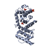
| ||||||||
| 2 | 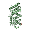
| ||||||||
| 3 |
| ||||||||
| Unit cell |
|
-Components
| #1: Protein | Mass: 30502.736 Da / Num. of mol.: 2 / Fragment: Actin-binding domain / Mutation: E254K Source method: isolated from a genetically manipulated source Source: (gene. exp.) Homo sapiens (human) / Gene: FLNA / Plasmid: pProEX HTb / Production host:  #2: Chemical | ChemComp-PO4 /   Mass: 94.971 Da / Num. of mol.: 4 / Source method: obtained synthetically / Formula: PO4 Mass: 94.971 Da / Num. of mol.: 4 / Source method: obtained synthetically / Formula: PO4#3: Water | ChemComp-HOH / |  Mass: 18.015 Da / Num. of mol.: 103 / Source method: isolated from a natural source / Formula: H2O Mass: 18.015 Da / Num. of mol.: 103 / Source method: isolated from a natural source / Formula: H2OHas protein modification | Y | |
|---|
-Experimental details
-Experiment
| Experiment | Method: X-RAY DIFFRACTION / Number of used crystals: 1 |
|---|
- Sample preparation
| Crystal | Density Matthews: 2.7 Å3/Da / Density % sol: 54.41 % |
|---|---|
| Crystal grow | Temperature: 293 K / Method: vapor diffusion, hanging drop / pH: 8.8 Details: 20% PEG 4000, 0.2M lithium sulfate, 0.1M Tris HCl, 5mM DTT, pH 8.8, VAPOR DIFFUSION, HANGING DROP, temperature 293K |
-Data collection
| Diffraction | Mean temperature: 120 K |
|---|---|
| Diffraction source | Source: ROTATING ANODE / Type: RIGAKU MICROMAX-007 / Wavelength: 1.5418 Å |
| Detector | Type: RIGAKU RAXIS IV / Detector: IMAGE PLATE / Date: Nov 15, 2006 / Details: osmic mirrors |
| Radiation | Monochromator: osmic mirrors / Protocol: SINGLE WAVELENGTH / Monochromatic (M) / Laue (L): M / Scattering type: x-ray |
| Radiation wavelength | Wavelength: 1.5418 Å / Relative weight: 1 |
| Reflection | Resolution: 2.3→57.64 Å / Num. obs: 29480 / % possible obs: 98 % / Redundancy: 4.2 % / Biso Wilson estimate: 33.25 Å2 / Rmerge(I) obs: 0.088 / Net I/σ(I): 12.3 / Num. measured all: 122444 |
| Reflection shell | Resolution: 2.3→2.42 Å / Redundancy: 4.1 % / Rmerge(I) obs: 0.53 / Mean I/σ(I) obs: 2.4 / Num. unique all: 2128 / % possible all: 93 |
- Processing
| Software |
| |||||||||||||||||||||||||
|---|---|---|---|---|---|---|---|---|---|---|---|---|---|---|---|---|---|---|---|---|---|---|---|---|---|---|
| Refinement | Method to determine structure: MOLECULAR REPLACEMENT Starting model: PDB ENTRY 1WKU Resolution: 2.3→43.62 Å / Cor.coef. Fo:Fc: 0.929 / Cor.coef. Fo:Fc free: 0.906 / Occupancy max: 1 / Occupancy min: 0 / SU B: 0.004 / SU ML: 0 / Cross valid method: THROUGHOUT / ESU R: 0.188 / ESU R Free: 0.223 / Stereochemistry target values: MAXIMUM LIKELIHOOD Details: HYDROGENS HAVE BEEN ADDED IN THE RIDING POSITIONS; PHENIX WAS ALSO USED FOR THE REFINEMENT
| |||||||||||||||||||||||||
| Solvent computation | Ion probe radii: 0.8 Å / Shrinkage radii: 0.8 Å / VDW probe radii: 1.4 Å / Solvent model: MASK | |||||||||||||||||||||||||
| Displacement parameters | Biso mean: 38.897 Å2
| |||||||||||||||||||||||||
| Refinement step | Cycle: LAST / Resolution: 2.3→43.62 Å
| |||||||||||||||||||||||||
| LS refinement shell | Resolution: 2.3→2.36 Å / Total num. of bins used: 20
|
