 Movie
Movie Controller

[English] 日本語
 Yorodumi
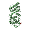
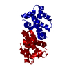
- PDB-3hor: Structure of the actin-binding domain of human filamin A (reduced) -
+ Open data

- Basic information
| Entry | Database: PDB / ID: 3hor | ||||||
|---|---|---|---|---|---|---|---|
| Title | Structure of the actin-binding domain of human filamin A (reduced) | ||||||
 Components Components | Filamin-A | ||||||
 Keywords Keywords | STRUCTURAL PROTEIN / Calponin homology domain / actin binding domain / Acetylation / Actin-binding / Alternative splicing / Cytoplasm / Cytoskeleton / Disease mutation / Phosphoprotein / Polymorphism | ||||||
| Function / homology |  Function and homology information Function and homology informationregulation of membrane repolarization during atrial cardiac muscle cell action potential / regulation of membrane repolarization during cardiac muscle cell action potential / establishment of Sertoli cell barrier / formation of radial glial scaffolds / Myb complex / adenylate cyclase-inhibiting dopamine receptor signaling pathway / positive regulation of integrin-mediated signaling pathway / blood coagulation, intrinsic pathway / OAS antiviral response / protein localization to bicellular tight junction ...regulation of membrane repolarization during atrial cardiac muscle cell action potential / regulation of membrane repolarization during cardiac muscle cell action potential / establishment of Sertoli cell barrier / formation of radial glial scaffolds / Myb complex / adenylate cyclase-inhibiting dopamine receptor signaling pathway / positive regulation of integrin-mediated signaling pathway / blood coagulation, intrinsic pathway / OAS antiviral response / protein localization to bicellular tight junction / actin crosslink formation / positive regulation of actin filament bundle assembly / positive regulation of neuron migration / tubulin deacetylation / megakaryocyte development / Cell-extracellular matrix interactions / positive regulation of platelet activation / positive regulation of potassium ion transmembrane transport / apical dendrite / Fc-gamma receptor I complex binding / positive regulation of neural precursor cell proliferation / protein localization to cell surface / negative regulation of transcription by RNA polymerase I / podosome / wound healing, spreading of cells / GP1b-IX-V activation signalling / SMAD binding / receptor clustering / cortical cytoskeleton / RHO GTPases activate PAKs / semaphorin-plexin signaling pathway / mitotic spindle assembly / cilium assembly / potassium channel regulator activity / release of sequestered calcium ion into cytosol / positive regulation of substrate adhesion-dependent cell spreading / regulation of cell migration / protein localization to plasma membrane / dendritic shaft / actin filament / establishment of protein localization / negative regulation of protein catabolic process / protein sequestering activity / cerebral cortex development / positive regulation of protein import into nucleus / platelet aggregation / mRNA transcription by RNA polymerase II / G protein-coupled receptor binding / small GTPase binding / kinase binding / Z disc / cell-cell junction / actin filament binding / actin cytoskeleton / Platelet degranulation / growth cone / actin cytoskeleton organization / GTPase binding / DNA-binding transcription factor binding / perikaryon / transmembrane transporter binding / positive regulation of canonical NF-kappaB signal transduction / postsynapse / protein stabilization / cadherin binding / focal adhesion / nucleolus / negative regulation of apoptotic process / perinuclear region of cytoplasm / glutamatergic synapse / protein homodimerization activity / RNA binding / extracellular exosome / extracellular region / membrane / nucleus / plasma membrane / cytosol / cytoplasm Similarity search - Function | ||||||
| Biological species |  Homo sapiens (human) Homo sapiens (human) | ||||||
| Method |  X-RAY DIFFRACTION / MOLECULAR REPLACEMENT / Resolution: 2.7 Å X-RAY DIFFRACTION / MOLECULAR REPLACEMENT / Resolution: 2.7 Å | ||||||
 Authors Authors | Clark, A.R. / Sawyer, G.M. / Robertson, S.P. / Sutherland-Smith, A.J. | ||||||
 Citation Citation | Journal: Hum.Mol.Genet. / Year: 2009 Title: Skeletal dysplasias due to filamin A mutations result from a gain-of-function mechanism distinct from allelic neurological disorders Authors: Clark, A.R. / Sawyer, G.M. / Robertson, S.P. / Sutherland-Smith, A.J. | ||||||
| History |
|
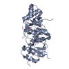
- Structure visualization
| Structure viewer | Molecule: MolmilJmol/JSmol |
|---|
- Downloads & links
-Download
| PDBx/mmCIF format | 3hor.cif.gz | 180.6 KB | Display | PDBx/mmCIF format |
|---|---|---|---|---|
| PDB format | pdb3hor.ent.gz | 143.1 KB | Display | PDB format |
| PDBx/mmJSON format | 3hor.json.gz | Tree view | PDBx/mmJSON format | |
| Others |  Other downloads Other downloads |
-Validation report
| Arichive directory | https://data.pdbj.org/pub/pdb/validation_reports/ho/3horftp://data.pdbj.org/pub/pdb/validation_reports/ho/3hor | HTTPS FTP |
|---|
-Related structure data
| Related structure data |  3hocC  3hopSC S: Starting model for refinement C: citing same article ( |
|---|---|
| Similar structure data |


-Links
 PDBj
PDBj


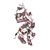
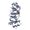
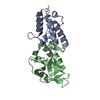
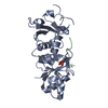
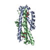
- Assembly
| Deposited unit | 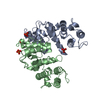
| |||||||||||||||||||||||||||||||||||||||||||||||||||||||||||||||||||||||
|---|---|---|---|---|---|---|---|---|---|---|---|---|---|---|---|---|---|---|---|---|---|---|---|---|---|---|---|---|---|---|---|---|---|---|---|---|---|---|---|---|---|---|---|---|---|---|---|---|---|---|---|---|---|---|---|---|---|---|---|---|---|---|---|---|---|---|---|---|---|---|---|---|
| 1 | 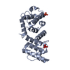
| |||||||||||||||||||||||||||||||||||||||||||||||||||||||||||||||||||||||
| 2 | 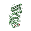
| |||||||||||||||||||||||||||||||||||||||||||||||||||||||||||||||||||||||
| Unit cell |
| |||||||||||||||||||||||||||||||||||||||||||||||||||||||||||||||||||||||
| Noncrystallographic symmetry (NCS) | NCS domain:
NCS domain segments: Ens-ID: 1
|