 ムービー
ムービー コントローラー


+ データを開く

- 基本情報
基本情報
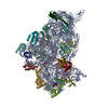
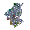
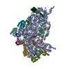
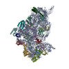
| 登録情報 | データベース: EMDB / ID: EMD-6710 | |||||||||
|---|---|---|---|---|---|---|---|---|---|---|
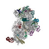
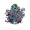
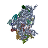
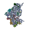
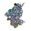
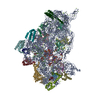
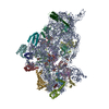
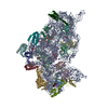
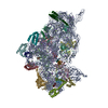
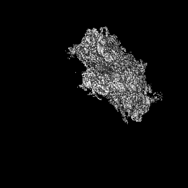
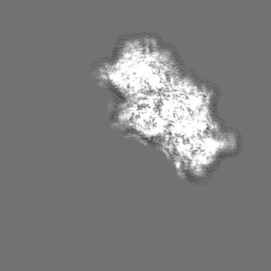
| タイトル | Structure of the 30S small subunit of chloroplast ribosome from spinach | |||||||||
 マップデータ マップデータ | ||||||||||
 試料 試料 |
| |||||||||
 キーワード キーワード | Cryo-EM / ribosome / chloroplast ribosome | |||||||||
| 機能・相同性 |  機能・相同性情報 機能・相同性情報chloroplast rRNA processing / plastid small ribosomal subunit / plastid translation / negative regulation of translational elongation / mitochondrial small ribosomal subunit / mitochondrial translation / chloroplast stroma / plastid / chloroplast thylakoid membrane / ribosomal small subunit binding ...chloroplast rRNA processing / plastid small ribosomal subunit / plastid translation / negative regulation of translational elongation / mitochondrial small ribosomal subunit / mitochondrial translation / chloroplast stroma / plastid / chloroplast thylakoid membrane / ribosomal small subunit binding / chloroplast / ribosomal small subunit assembly / ribosome biogenesis / ribosomal small subunit biogenesis / small ribosomal subunit / small ribosomal subunit rRNA binding / cytosolic small ribosomal subunit / rRNA binding / structural constituent of ribosome / ribosome / translation / ribonucleoprotein complex / response to antibiotic / mRNA binding / mitochondrion / RNA binding 類似検索 - 分子機能 | |||||||||
| 生物種 |  Spinacia oleracea (ホウレンソウ) Spinacia oleracea (ホウレンソウ) | |||||||||
| 手法 | 単粒子再構成法 / クライオ電子顕微鏡法 / 解像度: 3.7 Å | |||||||||
 データ登録者 データ登録者 | Ahmed T / Shi J | |||||||||
| 資金援助 |  シンガポール, 1件 シンガポール, 1件
| |||||||||
 引用 引用 | ジャーナル: Nucleic Acids Res / 年: 2017 タイトル: Unique localization of the plastid-specific ribosomal proteins in the chloroplast ribosome small subunit provides mechanistic insights into the chloroplastic translation. 著者: Tofayel Ahmed / Jian Shi / Shashi Bhushan / 要旨: Chloroplastic translation is mediated by a bacterial-type 70S chloroplast ribosome. During the evolution, chloroplast ribosomes have acquired five plastid-specific ribosomal proteins or PSRPs (cS22, ...Chloroplastic translation is mediated by a bacterial-type 70S chloroplast ribosome. During the evolution, chloroplast ribosomes have acquired five plastid-specific ribosomal proteins or PSRPs (cS22, cS23, bTHXc, cL37 and cL38) which have been suggested to play important regulatory roles in translation. However, their exact locations on the chloroplast ribosome remain elusive due to lack of a high-resolution structure, hindering our progress to understand their possible roles. Here we present a cryo-EM structure of the 70S chloroplast ribosome from spinach resolved to 3.4 Å and focus our discussion mainly on the architecture of the 30S small subunit (SSU) which is resolved to 3.7 Å. cS22 localizes at the SSU foot where it seems to compensate for the deletions in 16S rRNA. The mRNA exit site is highly remodeled due to the presence of cS23 suggesting an alternative mode of translation initiation. bTHXc is positioned at the SSU head and appears to stabilize the intersubunit bridge B1b during thermal fluctuations. The translation factor plastid pY binds to the SSU on the intersubunit side and interacts with the conserved nucleotide bases involved in decoding. Most of the intersubunit bridges are conserved compared to the bacteria, except for a new bridge involving uL2c and bS6c. | |||||||||
| 履歴 |
|
- 構造の表示
構造の表示
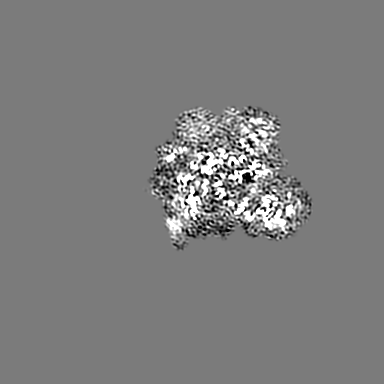
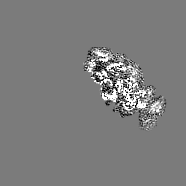
| ムービー |
ムービービューア |
|---|---|
| 構造ビューア | EMマップ: SurfViewMolmilJmol/JSmol |
| 添付画像 |

- ダウンロードとリンク
-EMDBアーカイブ
| マップデータ | emd_6710.map.gz | 9.8 MB | EMDBマップデータ形式 | |
|---|---|---|---|---|
| ヘッダ (付随情報) | emd-6710-v30.xmlemd-6710.xml | 38.2 KB 38.2 KB | 表示 表示 | EMDBヘッダ |
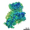
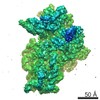
| 画像 |  emd_6710.png emd_6710.png | 81 KB | ||
| Filedesc metadata | emd-6710.cif.gz | 9.4 KB | ||
| アーカイブディレクトリ |  http://ftp.pdbj.org/pub/emdb/structures/EMD-6710ftp://ftp.pdbj.org/pub/emdb/structures/EMD-6710 http://ftp.pdbj.org/pub/emdb/structures/EMD-6710ftp://ftp.pdbj.org/pub/emdb/structures/EMD-6710 | HTTPS FTP |
-関連構造データ


-リンク
| EMDBのページ | EMDB (EBI/PDBe) / EMDataResource |
|---|---|
| 「今月の分子」の関連する項目 |


-マップ
| ファイル | ダウンロード / ファイル: emd_6710.map.gz / 形式: CCP4 / 大きさ: 216 MB / タイプ: IMAGE STORED AS FLOATING POINT NUMBER (4 BYTES) | ||||||||||||||||||||||||||||||||||||||||||||||||||||||||||||
|---|---|---|---|---|---|---|---|---|---|---|---|---|---|---|---|---|---|---|---|---|---|---|---|---|---|---|---|---|---|---|---|---|---|---|---|---|---|---|---|---|---|---|---|---|---|---|---|---|---|---|---|---|---|---|---|---|---|---|---|---|---|
| 投影像・断面図 | 画像のコントロール
画像は Spider により作成 | ||||||||||||||||||||||||||||||||||||||||||||||||||||||||||||
| ボクセルのサイズ | X=Y=Z: 1.05 Å | ||||||||||||||||||||||||||||||||||||||||||||||||||||||||||||
| 密度 |
| ||||||||||||||||||||||||||||||||||||||||||||||||||||||||||||
| 対称性 | 空間群: 1 | ||||||||||||||||||||||||||||||||||||||||||||||||||||||||||||
| 詳細 | EMDB XML:
CCP4マップ ヘッダ情報:
| ||||||||||||||||||||||||||||||||||||||||||||||||||||||||||||
 Z (Sec.)
 Y (Row.)
 X (Col.)


-添付データ
- 試料の構成要素
+全体 : 30S small subunit of chloroplast ribosome from spinach
+超分子 #1: 30S small subunit of chloroplast ribosome from spinach
+分子 #1: 30S ribosomal protein S2, chloroplastic
+分子 #2: 30S ribosomal protein S3, chloroplastic
+分子 #3: 30S ribosomal protein S5, chloroplastic
+分子 #4: 30S ribosomal protein S6 alpha, chloroplastic
+分子 #5: 30S ribosomal protein S7, chloroplastic
+分子 #6: 30S ribosomal protein S8, chloroplastic
+分子 #7: 30S ribosomal protein S9, chloroplastic
+分子 #8: protein S10
+分子 #9: 30S ribosomal protein S11, chloroplastic
+分子 #10: 30S ribosomal protein S12, chloroplastic
+分子 #11: 30S ribosomal protein S13, chloroplastic
+分子 #12: 30S ribosomal protein S15, chloroplastic
+分子 #13: 30S ribosomal protein S16, chloroplastic
+分子 #14: protein S17
+分子 #15: 30S ribosomal protein S18, chloroplastic
+分子 #16: 30S ribosomal protein S19 alpha, chloroplastic
+分子 #17: protein S20
+分子 #18: protein S21
+分子 #19: protein plastid pY
+分子 #21: protein cS23
+分子 #22: 30S ribosomal protein S4, chloroplastic
+分子 #23: protein cS22
+分子 #24: 30S ribosomal protein S14, chloroplastic
+分子 #25: protein bTHXc
+分子 #26: 30S ribosomal protein S1, chloroplastic
+分子 #20: 16S rRNA
-実験情報
-構造解析
| 手法 | クライオ電子顕微鏡法 |
|---|---|
 解析 解析 | 単粒子再構成法 |
| 試料の集合状態 | particle |
-試料調製
| 緩衝液 | pH: 7.6 詳細: 20 mM Tris HCl, pH 7.6, 100 mM KCl, 10 mM MgOAc, 100 mM sucrose, 7 mM 2-mercaptoethanol, 1 unit/ml RNase inhibitor, 0.1% protease inhibitor |
|---|---|
| グリッド | モデル: Quantifoil / 材質: COPPER / メッシュ: 300 / 支持フィルム - 材質: CARBON / 支持フィルム - トポロジー: CONTINUOUS / 支持フィルム - Film thickness: 2 / 前処理 - タイプ: GLOW DISCHARGE / 前処理 - 時間: 30 sec. |
| 凍結 | 凍結剤: ETHANE / チャンバー内湿度: 100 % / チャンバー内温度: 277 K / 装置: FEI VITROBOT MARK IV |
- 電子顕微鏡法
| 顕微鏡 | FEI TITAN KRIOS |
|---|---|
| 撮影 | フィルム・検出器のモデル: FEI FALCON II (4k x 4k) 検出モード: INTEGRATING / デジタル化 - サイズ - 横: 4096 pixel / デジタル化 - サイズ - 縦: 4096 pixel / デジタル化 - 画像ごとのフレーム数: 1-25 / 撮影したグリッド数: 1 / 実像数: 3161 / 平均電子線量: 1.5 e/Å2 |
| 電子線 | 加速電圧: 300 kV / 電子線源:  FIELD EMISSION GUN FIELD EMISSION GUN |
| 電子光学系 | C2レンズ絞り径: 100.0 µm / 最大 デフォーカス(補正後): 3.7 µm / 最小 デフォーカス(補正後): 0.4 µm / 倍率(補正後): 133333 / 照射モード: SPOT SCAN / 撮影モード: BRIGHT FIELD / Cs: 2.7 mm / 最大 デフォーカス(公称値): 3.7 µm / 最小 デフォーカス(公称値): 0.4 µm / 倍率(公称値): 75000 |
| 試料ステージ | 試料ホルダーモデル: FEI TITAN KRIOS AUTOGRID HOLDER ホルダー冷却材: NITROGEN |
| 実験機器 |  モデル: Titan Krios / 画像提供: FEI Company |
