+ データを開く
- 基本情報
基本情報
| 登録情報 | データベース: EMDB / ID: EMD-6343 | |||||||||
|---|---|---|---|---|---|---|---|---|---|---|
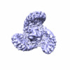
| タイトル | Cryo-EM study of a channel | |||||||||
 マップデータ マップデータ | Reconstruction of channel | |||||||||
 試料 試料 |
| |||||||||
 キーワード キーワード | Cryo-EM / single particle | |||||||||
| 機能・相同性 |  機能・相同性情報 機能・相同性情報mechanosensitive monoatomic cation channel activity / cuticular plate / positive regulation of cell-cell adhesion mediated by integrin / detection of mechanical stimulus / positive regulation of integrin activation / mechanosensitive monoatomic ion channel activity / stereocilium / positive regulation of myotube differentiation / lamellipodium membrane / monoatomic cation transport ...mechanosensitive monoatomic cation channel activity / cuticular plate / positive regulation of cell-cell adhesion mediated by integrin / detection of mechanical stimulus / positive regulation of integrin activation / mechanosensitive monoatomic ion channel activity / stereocilium / positive regulation of myotube differentiation / lamellipodium membrane / monoatomic cation transport / monoatomic cation channel activity / endoplasmic reticulum-Golgi intermediate compartment membrane / regulation of membrane potential / endoplasmic reticulum membrane / endoplasmic reticulum / identical protein binding / plasma membrane 類似検索 - 分子機能 | |||||||||
| 生物種 |  | |||||||||
| 手法 | 単粒子再構成法 / クライオ電子顕微鏡法 / ネガティブ染色法 / 解像度: 4.8 Å | |||||||||
 データ登録者 データ登録者 | Ge J / Li W / Zhao Q / Li N / Chen M / Zhi P / Li R / Gao N / Xiao B / Yang M | |||||||||
 引用 引用 |  ジャーナル: Nature / 年: 2015 ジャーナル: Nature / 年: 2015タイトル: Architecture of the mammalian mechanosensitive Piezo1 channel. 著者: Jingpeng Ge / Wanqiu Li / Qiancheng Zhao / Ningning Li / Maofei Chen / Peng Zhi / Ruochong Li / Ning Gao / Bailong Xiao / Maojun Yang /  要旨: Piezo proteins are evolutionarily conserved and functionally diverse mechanosensitive cation channels. However, the overall structural architecture and gating mechanisms of Piezo channels have ...Piezo proteins are evolutionarily conserved and functionally diverse mechanosensitive cation channels. However, the overall structural architecture and gating mechanisms of Piezo channels have remained unknown. Here we determine the cryo-electron microscopy structure of the full-length (2,547 amino acids) mouse Piezo1 (Piezo1) at a resolution of 4.8 Å. Piezo1 forms a trimeric propeller-like structure (about 900 kilodalton), with the extracellular domains resembling three distal blades and a central cap. The transmembrane region has 14 apparently resolved segments per subunit. These segments form three peripheral wings and a central pore module that encloses a potential ion-conducting pore. The rather flexible extracellular blade domains are connected to the central intracellular domain by three long beam-like structures. This trimeric architecture suggests that Piezo1 may use its peripheral regions as force sensors to gate the central ion-conducting pore. | |||||||||
| 履歴 |
|
- 構造の表示
構造の表示
| ムービー |
 ムービービューア ムービービューア |
|---|---|
| 構造ビューア | EMマップ:  SurfView SurfView Molmil Molmil Jmol/JSmol Jmol/JSmol |
| 添付画像 |
- ダウンロードとリンク
-EMDBアーカイブ
| マップデータ |  emd_6343.map.gz emd_6343.map.gz | 47.8 MB |  EMDBマップデータ形式 EMDBマップデータ形式 | |
|---|---|---|---|---|
| ヘッダ (付随情報) |  emd-6343-v30.xml emd-6343-v30.xml emd-6343.xml emd-6343.xml | 9.7 KB 9.7 KB | 表示 表示 |  EMDBヘッダ EMDBヘッダ |
| 画像 |  400_6343.gif 400_6343.gif 80_6343.gif 80_6343.gif | 35.7 KB 2.8 KB | ||
| アーカイブディレクトリ |  http://ftp.pdbj.org/pub/emdb/structures/EMD-6343 http://ftp.pdbj.org/pub/emdb/structures/EMD-6343 ftp://ftp.pdbj.org/pub/emdb/structures/EMD-6343 ftp://ftp.pdbj.org/pub/emdb/structures/EMD-6343 | HTTPS FTP |
-検証レポート
| 文書・要旨 |  emd_6343_validation.pdf.gz emd_6343_validation.pdf.gz | 358.8 KB | 表示 |  EMDB検証レポート EMDB検証レポート |
|---|---|---|---|---|
| 文書・詳細版 |  emd_6343_full_validation.pdf.gz emd_6343_full_validation.pdf.gz | 358.4 KB | 表示 | |
| XML形式データ |  emd_6343_validation.xml.gz emd_6343_validation.xml.gz | 5.8 KB | 表示 | |
| アーカイブディレクトリ |  https://ftp.pdbj.org/pub/emdb/validation_reports/EMD-6343 https://ftp.pdbj.org/pub/emdb/validation_reports/EMD-6343 ftp://ftp.pdbj.org/pub/emdb/validation_reports/EMD-6343 ftp://ftp.pdbj.org/pub/emdb/validation_reports/EMD-6343 | HTTPS FTP |
-関連構造データ
- リンク
リンク
| EMDBのページ |  EMDB (EBI/PDBe) / EMDB (EBI/PDBe) /  EMDataResource EMDataResource |
|---|---|
| 「今月の分子」の関連する項目 |
- マップ
マップ
| ファイル |  ダウンロード / ファイル: emd_6343.map.gz / 形式: CCP4 / 大きさ: 51.5 MB / タイプ: IMAGE STORED AS FLOATING POINT NUMBER (4 BYTES) ダウンロード / ファイル: emd_6343.map.gz / 形式: CCP4 / 大きさ: 51.5 MB / タイプ: IMAGE STORED AS FLOATING POINT NUMBER (4 BYTES) | ||||||||||||||||||||||||||||||||||||||||||||||||||||||||||||
|---|---|---|---|---|---|---|---|---|---|---|---|---|---|---|---|---|---|---|---|---|---|---|---|---|---|---|---|---|---|---|---|---|---|---|---|---|---|---|---|---|---|---|---|---|---|---|---|---|---|---|---|---|---|---|---|---|---|---|---|---|---|
| 注釈 | Reconstruction of channel | ||||||||||||||||||||||||||||||||||||||||||||||||||||||||||||
| 投影像・断面図 | 画像のコントロール
画像は Spider により作成 | ||||||||||||||||||||||||||||||||||||||||||||||||||||||||||||
| ボクセルのサイズ | X=Y=Z: 1.32 Å | ||||||||||||||||||||||||||||||||||||||||||||||||||||||||||||
| 密度 |
| ||||||||||||||||||||||||||||||||||||||||||||||||||||||||||||
| 対称性 | 空間群: 1 | ||||||||||||||||||||||||||||||||||||||||||||||||||||||||||||
| 詳細 | EMDB XML:
CCP4マップ ヘッダ情報:
| ||||||||||||||||||||||||||||||||||||||||||||||||||||||||||||
-添付データ
- 試料の構成要素
-全体 : Piezo-type mechanosensitive ion channel component 1
| 全体 | 名称: Piezo-type mechanosensitive ion channel component 1 |
|---|---|
| 要素 |
|
-超分子 #1000: Piezo-type mechanosensitive ion channel component 1
| 超分子 | 名称: Piezo-type mechanosensitive ion channel component 1 / タイプ: sample / ID: 1000 / Number unique components: 1 |
|---|---|
| 分子量 | 理論値: 900 KDa |
-分子 #1: Piezo-type mechanosensitive ion channel component 1
| 分子 | 名称: Piezo-type mechanosensitive ion channel component 1 / タイプ: protein_or_peptide / ID: 1 / コピー数: 3 / 組換発現: Yes |
|---|---|
| 由来(天然) | 生物種:  |
| 組換発現 | 生物種:  Homo sapiens (ヒト) / 組換細胞: HEK 293T / 組換プラスミド: pCDNA3.1(-) Homo sapiens (ヒト) / 組換細胞: HEK 293T / 組換プラスミド: pCDNA3.1(-) |
| 配列 | UniProtKB: Piezo-type mechanosensitive ion channel component 1 |
-実験情報
-構造解析
| 手法 | ネガティブ染色法, クライオ電子顕微鏡法 |
|---|---|
 解析 解析 | 単粒子再構成法 |
| 試料の集合状態 | particle |
- 試料調製
試料調製
| 緩衝液 | pH: 7.2 詳細: 25 mM NaPIPES, 140 mM NaCl, 2 mM DTT and 0.026% (w/v) C12E10 |
|---|---|
| 染色 | タイプ: NEGATIVE 詳細: Grids with adsorbed protein floated on 2% w/v uranyl acetate for 30 seconds |
| 凍結 | 凍結剤: ETHANE / チャンバー内湿度: 100 % / 装置: FEI VITROBOT MARK IV |
- 電子顕微鏡法
| 顕微鏡 | FEI TITAN KRIOS |
|---|---|
| 日付 | 2014年4月25日 |
| 撮影 | カテゴリ: CCD / フィルム・検出器のモデル: GATAN K2 (4k x 4k) / デジタル化 - サンプリング間隔: 4 µm / 実像数: 2164 / 平均電子線量: 16 e/Å2 詳細: Every image is the average of 14 frames recorded by the direct electron detector |
| 電子線 | 加速電圧: 300 kV / 電子線源:  FIELD EMISSION GUN FIELD EMISSION GUN |
| 電子光学系 | 照射モード: FLOOD BEAM / 撮影モード: BRIGHT FIELD / 倍率(公称値): 22500 |
| 試料ステージ | 試料ホルダーモデル: FEI TITAN KRIOS AUTOGRID HOLDER |
| 実験機器 |  モデル: Titan Krios / 画像提供: FEI Company |
- 画像解析
画像解析
| CTF補正 | 詳細: CTFFIND |
|---|---|
| 最終 再構成 | 解像度のタイプ: BY AUTHOR / 解像度: 4.8 Å / 解像度の算出法: OTHER / ソフトウェア - 名称: RELION / 使用した粒子像数: 30021 |
 ムービー
ムービー コントローラー


 Z (Sec.)
 Y (Row.)
 X (Col.)


