 Movie
Movie Controller


+ Open data

- Basic information
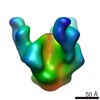
| Entry | Database: PDB / ID: 3jac | ||||||
|---|---|---|---|---|---|---|---|
| Title | Cryo-EM study of a channel | ||||||
 Components Components | Piezo-type mechanosensitive ion channel component 1 | ||||||
 Keywords Keywords | METAL TRANSPORT / Cryo-EM / single particle | ||||||
| Function / homology |  Function and homology information Function and homology informationmechanosensitive monoatomic cation channel activity / cuticular plate / positive regulation of integrin activation / detection of mechanical stimulus / positive regulation of cell-cell adhesion mediated by integrin / mechanosensitive monoatomic ion channel activity / stereocilium / positive regulation of myotube differentiation / lamellipodium membrane / monoatomic cation transport ...mechanosensitive monoatomic cation channel activity / cuticular plate / positive regulation of integrin activation / detection of mechanical stimulus / positive regulation of cell-cell adhesion mediated by integrin / mechanosensitive monoatomic ion channel activity / stereocilium / positive regulation of myotube differentiation / lamellipodium membrane / monoatomic cation transport / monoatomic cation channel activity / endoplasmic reticulum-Golgi intermediate compartment membrane / regulation of membrane potential / cellular response to mechanical stimulus / endoplasmic reticulum membrane / endoplasmic reticulum / identical protein binding / plasma membrane Similarity search - Function | ||||||
| Biological species |  | ||||||
| Method | ELECTRON MICROSCOPY / single particle reconstruction / negative staining / cryo EM / Resolution: 4.8 Å | ||||||
 Authors Authors | Ge, J. / Li, W. / Zhao, Q. / Li, N. / Xiao, B. / Gao, N. / Yang, M. | ||||||
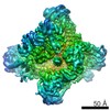
 Citation Citation | Journal: Nature / Year: 2015 Title: Architecture of the mammalian mechanosensitive Piezo1 channel. Authors: Jingpeng Ge / Wanqiu Li / Qiancheng Zhao / Ningning Li / Maofei Chen / Peng Zhi / Ruochong Li / Ning Gao / Bailong Xiao / Maojun Yang /  Abstract: Piezo proteins are evolutionarily conserved and functionally diverse mechanosensitive cation channels. However, the overall structural architecture and gating mechanisms of Piezo channels have ...Piezo proteins are evolutionarily conserved and functionally diverse mechanosensitive cation channels. However, the overall structural architecture and gating mechanisms of Piezo channels have remained unknown. Here we determine the cryo-electron microscopy structure of the full-length (2,547 amino acids) mouse Piezo1 (Piezo1) at a resolution of 4.8 Å. Piezo1 forms a trimeric propeller-like structure (about 900 kilodalton), with the extracellular domains resembling three distal blades and a central cap. The transmembrane region has 14 apparently resolved segments per subunit. These segments form three peripheral wings and a central pore module that encloses a potential ion-conducting pore. The rather flexible extracellular blade domains are connected to the central intracellular domain by three long beam-like structures. This trimeric architecture suggests that Piezo1 may use its peripheral regions as force sensors to gate the central ion-conducting pore. | ||||||
| History |
|
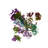
- Structure visualization
| Movie |
Movie viewer |
|---|---|
| Structure viewer | Molecule: MolmilJmol/JSmol |
 UCSF Chimera
- Downloads & links
-Download
| PDBx/mmCIF format | 3jac.cif.gz | 463.2 KB | Display | PDBx/mmCIF format |
|---|---|---|---|---|
| PDB format | pdb3jac.ent.gz | 303.6 KB | Display | PDB format |
| PDBx/mmJSON format | 3jac.json.gz | Tree view | PDBx/mmJSON format | |
| Others |  Other downloads Other downloads |
-Validation report
| Arichive directory | https://data.pdbj.org/pub/pdb/validation_reports/ja/3jacftp://data.pdbj.org/pub/pdb/validation_reports/ja/3jac | HTTPS FTP |
|---|
-Related structure data
| Related structure data | 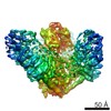 6343MC  4raxC M: map data used to model this data C: citing same article ( |
|---|---|
| Similar structure data |

-Links
 PDBj
PDBj
- Assembly
| Deposited unit | 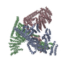
|
|---|---|
| 1 |
|
-Components
| #1: Protein | Mass: 234834.109 Da / Num. of mol.: 3 Source method: isolated from a genetically manipulated source Source: (gene. exp.)  Homo sapiens (human) / References: UniProt: E2JF22 Homo sapiens (human) / References: UniProt: E2JF22Has protein modification | Y | Sequence details | THE COMPLETE SEQUENCE OF THE PROTEIN IS BASED ON THE ENTIRE SEQUNCE OF UNIPROTKB E2JF22 (PIEZ1_ ...THE COMPLETE SEQUENCE OF THE PROTEIN IS BASED ON THE ENTIRE SEQUNCE OF UNIPROTKB E2JF22 (PIEZ1_MOUSE), WITH C-TERMINAL EXPRESSION | |
|---|