Movie
Movie Controller


+ Open data

- Basic information
| Entry | Database: EMDB / ID: EMD-2640 | |||||||||
|---|---|---|---|---|---|---|---|---|---|---|
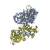
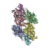
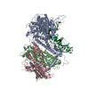
| Title | Negative-stain electron microscopy of the human GINS complex | |||||||||
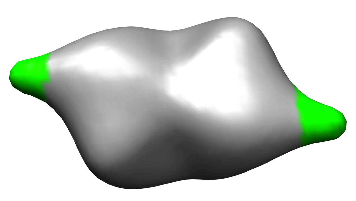
 Map data Map data | Reconstruction of the human GINS complex | |||||||||
 Sample Sample |
| |||||||||
 Keywords Keywords | DNA replication / CMG / GINS complex / Psf1 B domain | |||||||||
| Biological species |  Homo sapiens (human) Homo sapiens (human) | |||||||||
| Method | single particle reconstruction / negative staining / Resolution: 30.0 Å | |||||||||
 Authors Authors | Carroni M / De March M / Krastanova I / Medagli B / Taylor IK / Brick P / Amenitsch H / Patwardhan A / Onesti S | |||||||||
 Citation Citation | Journal: Sci Rep / Year: 2017 Title: New insights into the GINS complex explain the controversy between existing structural models. Authors: Marta Carroni / Matteo De March / Barbara Medagli / Ivet Krastanova / Ian A Taylor / Heinz Amenitsch / Hiroyuchi Araki / Francesca M Pisani / Ardan Patwardhan / Silvia Onesti /     Abstract: GINS is a key component of eukaryotic replicative forks and is composed of four subunits (Sld5, Psf1, Psf2, Psf3). To explain the discrepancy between structural data from crystallography and electron ...GINS is a key component of eukaryotic replicative forks and is composed of four subunits (Sld5, Psf1, Psf2, Psf3). To explain the discrepancy between structural data from crystallography and electron microscopy (EM), we show that GINS is a compact tetramer in solution as observed in crystal structures, but also forms a double-tetrameric population, detectable by EM. This may represent an intermediate step towards the assembly of two replicative helicase complexes at origins, moving in opposite directions within the replication bubble. Reconstruction of the double-tetrameric form, combined with small-angle X-ray scattering data, allows the localisation of the B domain of the Psf1 subunit in the free GINS complex, which was not visible in previous studies and is essential for the formation of a functional replication fork. | |||||||||
| History |
|
- Structure visualization
| Movie |
 Movie viewer Movie viewer |
|---|---|
| Structure viewer | EM map: SurfViewMolmilJmol/JSmol |
| Supplemental images |
- Downloads & links
-EMDB archive
| Map data | emd_2640.map.gz | 96.6 KB | EMDB map data format | |
|---|---|---|---|---|
| Header (meta data) | emd-2640-v30.xmlemd-2640.xml | 10.4 KB 10.4 KB | Display Display | EMDB header |
| Images |  emd_2640.png emd_2640.png | 342.3 KB | ||
| Archive directory |  http://ftp.pdbj.org/pub/emdb/structures/EMD-2640ftp://ftp.pdbj.org/pub/emdb/structures/EMD-2640 http://ftp.pdbj.org/pub/emdb/structures/EMD-2640ftp://ftp.pdbj.org/pub/emdb/structures/EMD-2640 | HTTPS FTP |
-Validation report
| Summary document | emd_2640_validation.pdf.gz | 186.7 KB | Display | EMDB validaton report |
|---|---|---|---|---|
| Full document | emd_2640_full_validation.pdf.gz | 185.9 KB | Display | |
| Data in XML | emd_2640_validation.xml.gz | 4.4 KB | Display | |
| Arichive directory | https://ftp.pdbj.org/pub/emdb/validation_reports/EMD-2640ftp://ftp.pdbj.org/pub/emdb/validation_reports/EMD-2640 | HTTPS FTP |
-Related structure data
| Similar structure data |
|---|


-Links
| EMDB pages | EMDB (EBI/PDBe) / EMDataResource |
|---|
-Map
| File | Download / File: emd_2640.map.gz / Format: CCP4 / Size: 1.9 MB / Type: IMAGE STORED AS FLOATING POINT NUMBER (4 BYTES) | ||||||||||||||||||||||||||||||||||||||||||||||||||||||||||||
|---|---|---|---|---|---|---|---|---|---|---|---|---|---|---|---|---|---|---|---|---|---|---|---|---|---|---|---|---|---|---|---|---|---|---|---|---|---|---|---|---|---|---|---|---|---|---|---|---|---|---|---|---|---|---|---|---|---|---|---|---|---|
| Annotation | Reconstruction of the human GINS complex | ||||||||||||||||||||||||||||||||||||||||||||||||||||||||||||
| Projections & slices | Image control
Images are generated by Spider. | ||||||||||||||||||||||||||||||||||||||||||||||||||||||||||||
| Voxel size | X=Y=Z: 4 Å | ||||||||||||||||||||||||||||||||||||||||||||||||||||||||||||
| Density |
| ||||||||||||||||||||||||||||||||||||||||||||||||||||||||||||
| Symmetry | Space group: 1 | ||||||||||||||||||||||||||||||||||||||||||||||||||||||||||||
| Details | EMDB XML:
CCP4 map header:
| ||||||||||||||||||||||||||||||||||||||||||||||||||||||||||||
 Z (Sec.)
 Y (Row.)
 X (Col.)


-Supplemental data
- Sample components
-Entire : Human GINS complex
| Entire | Name: Human GINS complex |
|---|---|
| Components |
|
-Supramolecule #1000: Human GINS complex
| Supramolecule | Name: Human GINS complex / type: sample / ID: 1000 Details: The sample was in equilibrium with a monomeric form (single GINS tetramer) Oligomeric state: dimer of human GINS tetramers / Number unique components: 1 |
|---|---|
| Molecular weight | Experimental: 200 KDa / Theoretical: 200 KDa Method: SEC-MALLS (size-exclusion chromatography coupled with multi angle laser light scattering) |
-Macromolecule #1: GINS
| Macromolecule | Name: GINS / type: protein_or_peptide / ID: 1 / Number of copies: 2 / Oligomeric state: dimer / Recombinant expression: Yes |
|---|---|
| Source (natural) | Organism: Homo sapiens (human) / synonym: Human / Tissue: all / Cell: all / Organelle: nucleus / Location in cell: nucleus |
| Molecular weight | Experimental: 100 KDa / Theoretical: 100 KDa |
| Recombinant expression | Organism:  |
-Experimental details
-Structure determination
| Method | negative staining |
|---|---|
 Processing Processing | single particle reconstruction |
| Aggregation state | particle |
-Sample preparation
| Concentration | 0.05 mg/mL |
|---|---|
| Buffer | pH: 7.8 / Details: 20 mM TrisHCl pH 7.8, 150 mM NaCl, 0.5 mM TCEP |
| Staining | Type: NEGATIVE Details: Protein adsorbed on carbon coated grids, stained with potassium phospho tungstate pH 7.0 for 4 minutes. |
| Vitrification | Cryogen name: NONE / Instrument: OTHER |
- Electron microscopy
| Microscope | FEI/PHILIPS CM200FEG |
|---|---|
| Alignment procedure | Legacy - Astigmatism: Objective lens astigmatism was corrected at 150,000 times magnification |
| Date | Dec 20, 2009 |
| Image recording | Category: CCD / Film or detector model: TVIPS TEMCAM-F416 (4k x 4k) / Average electron dose: 20 e/Å2 |
| Electron beam | Acceleration voltage: 200 kV / Electron source:  FIELD EMISSION GUN FIELD EMISSION GUN |
| Electron optics | Calibrated magnification: 50000 / Illumination mode: FLOOD BEAM / Imaging mode: BRIGHT FIELD / Cs: 1.2 mm / Nominal defocus max: 3.0 µm / Nominal defocus min: 1.0 µm / Nominal magnification: 50000 |
| Sample stage | Specimen holder model: SIDE ENTRY, EUCENTRIC |
-Image processing
| CTF correction | Details: entire frame |
|---|---|
| Final reconstruction | Applied symmetry - Point group: C2 (2 fold cyclic) / Algorithm: OTHER / Resolution.type: BY AUTHOR / Resolution: 30.0 Å / Resolution method: OTHER / Software - Name: IMAGIC, Spider / Number images used: 14390 |
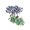
-Atomic model buiding 1
| Initial model | PDB ID: Chain - #0 - Chain ID: E / Chain - #1 - Chain ID: F / Chain - #2 - Chain ID: G / Chain - #3 - Chain ID: H / Chain - #4 - Chain ID: I / Chain - #5 - Chain ID: J / Chain - #6 - Chain ID: K / Chain - #7 - Chain ID: L |
|---|---|
| Software | Name: Chimera |
| Details | A dimer of tetramers observed in the crystal was fitted. |
| Refinement | Space: REAL / Protocol: RIGID BODY FIT |