National Institutes of Health/National Institute of General Medical Sciences (NIH/NIGMS)
R01GM116942
United States
National Institutes of Health/National Institute of General Medical Sciences (NIH/NIGMS)
R35GM136258
United States
National Institutes of Health/National Institute of General Medical Sciences (NIH/NIGMS)
R01GM097272
United States
National Institutes of Health/National Institute on Minority Health and Health Disparities (NIH/NIMHD)
R01HD095296
United States
National Institutes of Health/National Institute of General Medical Sciences (NIH/NIGMS)
R01GM124559
United States
National Institutes of Health/National Institute of General Medical Sciences (NIH/NIGMS)
R01GM124559
United States
National Science Foundation (NSF, United States)
DBI-1661380
United States
National Science Foundation (NSF, United States)
DMR-1719875
United States
Medical Research Council (MRC, United Kingdom)
MRC_UP_1201/10
United Kingdom
National Science Foundation (NSF, United States)
DGE-1650441
United States
Citation
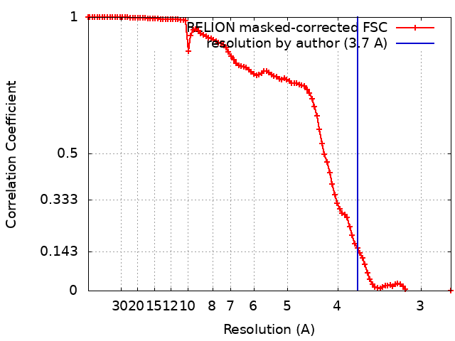
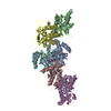
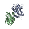
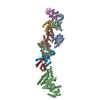
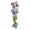
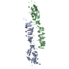
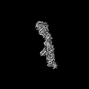
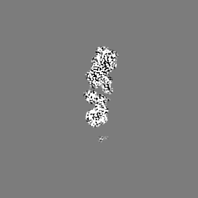
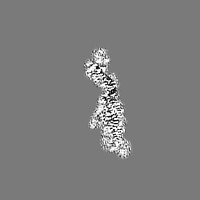
Journal: EMBO J / Year: 2021 Title: Structural basis of TRAPPIII-mediated Rab1 activation. Authors: Aaron Mn Joiner / Ben P Phillips / Kumar Yugandhar / Ethan J Sanford / Marcus B Smolka / Haiyuan Yu / Elizabeth A Miller / J Christopher Fromme / Abstract: The GTPase Rab1 is a master regulator of the early secretory pathway and is critical for autophagy. Rab1 activation is controlled by its guanine nucleotide exchange factor, the multisubunit TRAPPIII ...The GTPase Rab1 is a master regulator of the early secretory pathway and is critical for autophagy. Rab1 activation is controlled by its guanine nucleotide exchange factor, the multisubunit TRAPPIII complex. Here, we report the 3.7 Å cryo-EM structure of the Saccharomyces cerevisiae TRAPPIII complex bound to its substrate Rab1/Ypt1. The structure reveals the binding site for the Rab1/Ypt1 hypervariable domain, leading to a model for how the complex interacts with membranes during the activation reaction. We determined that stable membrane binding by the TRAPPIII complex is required for robust activation of Rab1/Ypt1 in vitro and in vivo, and is mediated by a conserved amphipathic α-helix within the regulatory Trs85 subunit. Our results show that the Trs85 subunit serves as a membrane anchor, via its amphipathic helix, for the entire TRAPPIII complex. These findings provide a structural understanding of Rab activation on organelle and vesicle membranes.
History
Deposition
Nov 3, 2020
-
Header (metadata) release
Jun 2, 2021
-
Map release
Jun 2, 2021
-
Update
Oct 16, 2024
-
Current status
Oct 16, 2024
Processing site: RCSB / Status: Released
-
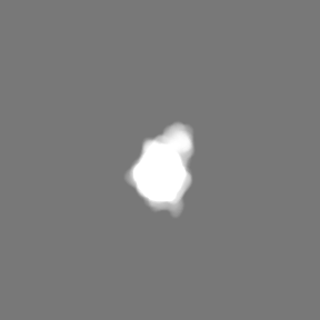
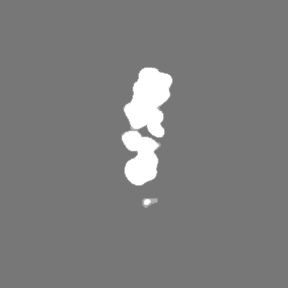
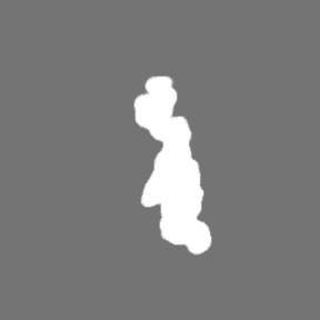
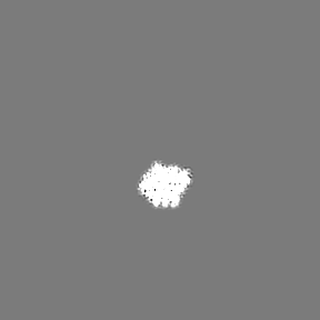
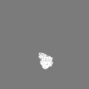
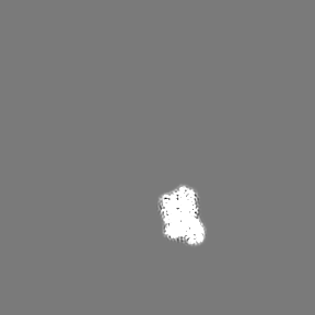
Structure visualization
Movie
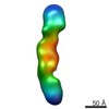
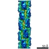
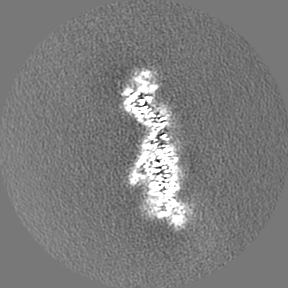
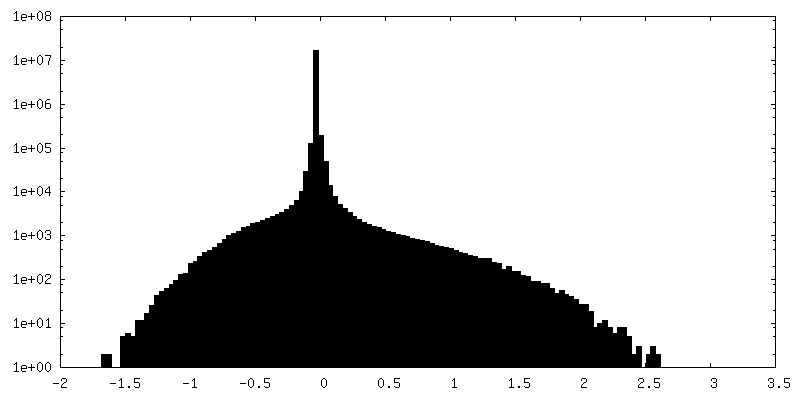
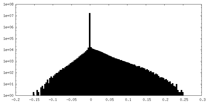
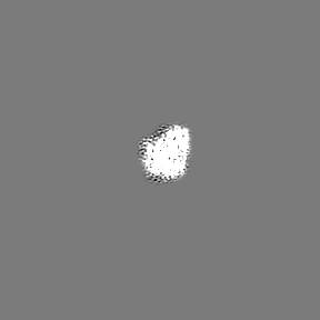
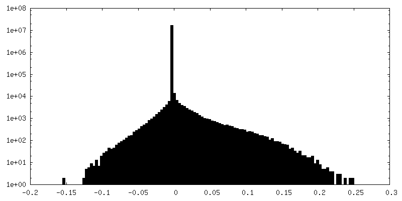
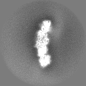
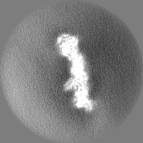
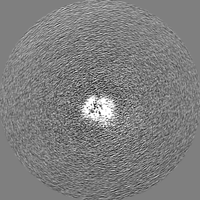
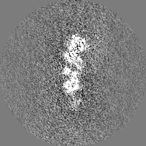
Surface view with section colored by density value
Model: UltrAuFoil R1.2/1.3 / Material: GOLD / Mesh: 300 / Support film - Material: GOLD / Support film - topology: HOLEY / Support film - Film thickness: 50 / Pretreatment - Type: GLOW DISCHARGE
Vitrification
Cryogen name: ETHANE / Chamber humidity: 100 % / Chamber temperature: 277 K / Instrument: FEI VITROBOT MARK IV Details: Either 0.02% Tween-20 or 0.025% amphipol A8-35 was added before application of the sample to the grid..
-
Electron microscopy #1
Microscopy ID
1
Microscope
FEI TALOS ARCTICA
Specialist optics
Energy filter - Name: GIF Bioquantum / Energy filter - Slit width: 20 eV
Image recording
Image recording ID: 1 / Film or detector model: GATAN K3 BIOQUANTUM (6k x 4k) / Average electron dose: 50.0 e/Å2
Electron beam
Acceleration voltage: 200 kV / Electron source: FIELD EMISSION GUN
Electron optics
Illumination mode: FLOOD BEAM / Imaging mode: BRIGHT FIELD
Experimental equipment
Model: Talos Arctica / Image courtesy: FEI Company
-
Electron microscopy #1~
Microscopy ID
1
Microscope
FEI TITAN KRIOS
Image recording
Image recording ID: 2 / Film or detector model: FEI FALCON III (4k x 4k) / Detector mode: COUNTING / Average electron dose: 20.0 e/Å2
Electron beam
Acceleration voltage: 300 kV / Electron source: FIELD EMISSION GUN
Electron optics
Illumination mode: FLOOD BEAM / Imaging mode: BRIGHT FIELD
Experimental equipment
Model: Titan Krios / Image courtesy: FEI Company
-
Electron microscopy #1~~
Microscopy ID
1
Microscope
FEI TITAN KRIOS
Specialist optics
Energy filter - Name: GIF Quantum LS / Energy filter - Slit width: 20 eV
Image recording
Image recording ID: 3 / Film or detector model: GATAN K2 SUMMIT (4k x 4k) / Detector mode: SUPER-RESOLUTION / Average electron dose: 50.0 e/Å2
Electron beam
Acceleration voltage: 300 kV / Electron source: FIELD EMISSION GUN
Electron optics
Illumination mode: FLOOD BEAM / Imaging mode: BRIGHT FIELD
In the structure databanks used in Yorodumi, some data are registered as the other names, "COVID-19 virus" and "2019-nCoV". Here are the details of the virus and the list of structure data.
Jan 31, 2019. EMDB accession codes are about to change! (news from PDBe EMDB page)
EMDB accession codes are about to change! (news from PDBe EMDB page)
The allocation of 4 digits for EMDB accession codes will soon come to an end. Whilst these codes will remain in use, new EMDB accession codes will include an additional digit and will expand incrementally as the available range of codes is exhausted. The current 4-digit format prefixed with “EMD-” (i.e. EMD-XXXX) will advance to a 5-digit format (i.e. EMD-XXXXX), and so on. It is currently estimated that the 4-digit codes will be depleted around Spring 2019, at which point the 5-digit format will come into force.
The EM Navigator/Yorodumi systems omit the EMD- prefix.
Related info.:Q: What is EMD? / ID/Accession-code notation in Yorodumi/EM Navigator
Yorodumi is a browser for structure data from EMDB, PDB, SASBDB, etc.
This page is also the successor to EM Navigator detail page, and also detail information page/front-end page for Omokage search.
The word "yorodu" (or yorozu) is an old Japanese word meaning "ten thousand". "mi" (miru) is to see.
Related info.:EMDB / PDB / SASBDB / Comparison of 3 databanks / Yorodumi Search / Aug 31, 2016. New EM Navigator & Yorodumi / Yorodumi Papers / Jmol/JSmol / Function and homology information / Changes in new EM Navigator and Yorodumi
 Movie
Movie Controller


 Open data
 Basic information
 Map data
 Sample
 Keywords
 Function and homology information
 Authors
 United States,
  United Kingdom, 10 items
  Citation
 Structure visualization
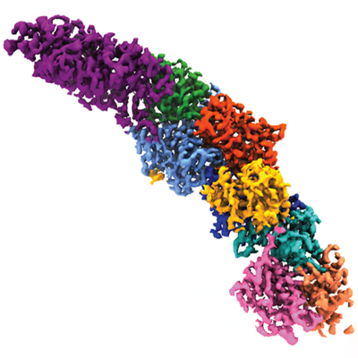
 Downloads & links
 emd_22928.png
 http://ftp.pdbj.org/pub/emdb/structures/EMD-22928
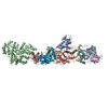


 Z (Sec.)
 Y (Row.)
 X (Col.)


 Sample components

 Processing
 Electron microscopy #1
 FIELD EMISSION GUN