 Movie
Movie Controller

[English] 日本語
 Yorodumi
- PDB-3cue: Crystal structure of a TRAPP subassembly activating the Rab Ypt1p -
+ Open data

- Basic information
| Entry | Database: PDB / ID: 3cue | ||||||
|---|---|---|---|---|---|---|---|
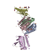
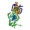
| Title | Crystal structure of a TRAPP subassembly activating the Rab Ypt1p | ||||||
 Components Components |
| ||||||
 Keywords Keywords | PROTEIN TRANSPORT / membrane traffic / GEF / tethering complex / Rab activation / guanine nucleotide exchange factor / Endoplasmic reticulum / ER-Golgi transport / Golgi apparatus / Transport / Lipoprotein / Palmitate / GTP-binding / Nucleotide-binding / Phosphoprotein / Prenylation | ||||||
| Function / homology |  Function and homology information Function and homology informationGolgi Cisternae Pericentriolar Stack Reorganization / : / pre-mRNA catabolic process / regulation of endoplasmic reticulum unfolded protein response / TRAPPI protein complex / RAB geranylgeranylation / TRAPPII protein complex / Golgi vesicle budding / RAB GEFs exchange GTP for GDP on RABs / TRAPPIII protein complex ...Golgi Cisternae Pericentriolar Stack Reorganization / : / pre-mRNA catabolic process / regulation of endoplasmic reticulum unfolded protein response / TRAPPI protein complex / RAB geranylgeranylation / TRAPPII protein complex / Golgi vesicle budding / RAB GEFs exchange GTP for GDP on RABs / TRAPPIII protein complex / TRAPP complex / early endosome to Golgi transport / COPI-dependent Golgi-to-ER retrograde traffic / COPII-coated vesicle budding / COPI-mediated anterograde transport / cytoplasm to vacuole targeting by the Cvt pathway / COPII-mediated vesicle transport / SNARE complex disassembly / phagophore assembly site membrane / protein localization to phagophore assembly site / cis-Golgi network membrane / cis-Golgi network / intra-Golgi vesicle-mediated transport / endocytic recycling / retrograde vesicle-mediated transport, Golgi to endoplasmic reticulum / Golgi stack / phagophore assembly site / retrograde transport, endosome to Golgi / reticulophagy / cellular response to nitrogen starvation / SNARE complex assembly / chromosome organization / endoplasmic reticulum to Golgi vesicle-mediated transport / autophagosome assembly / Neutrophil degranulation / endomembrane system / SNARE binding / macroautophagy / intracellular protein transport / cytoplasmic vesicle / Golgi membrane / GTPase activity / endoplasmic reticulum membrane / GTP binding / endoplasmic reticulum / mitochondrion / cytosol Similarity search - Function | ||||||
| Biological species |  | ||||||
| Method |  X-RAY DIFFRACTION / SYNCHROTRON / MAD / Resolution: 3.7 Å X-RAY DIFFRACTION / SYNCHROTRON / MAD / Resolution: 3.7 Å | ||||||
 Authors Authors | Cai, Y. / Reinisch, K.M. | ||||||
 Citation Citation | Journal: Cell(Cambridge,Mass.) / Year: 2008 Title: The structural basis for activation of the Rab Ypt1p by the TRAPP membrane-tethering complexes. Authors: Cai, Y. / Chin, H.F. / Lazarova, D. / Menon, S. / Fu, C. / Cai, H. / Sclafani, A. / Rodgers, D.W. / De La Cruz, E.M. / Ferro-Novick, S. / Reinisch, K.M. | ||||||
| History |
|
- Structure visualization
| Structure viewer | Molecule: MolmilJmol/JSmol |
|---|
- Downloads & links
-Download
| PDBx/mmCIF format | 3cue.cif.gz | 824.3 KB | Display | PDBx/mmCIF format |
|---|---|---|---|---|
| PDB format | pdb3cue.ent.gz | 664.2 KB | Display | PDB format |
| PDBx/mmJSON format | 3cue.json.gz | Tree view | PDBx/mmJSON format | |
| Others |  Other downloads Other downloads |
-Validation report
| Arichive directory | https://data.pdbj.org/pub/pdb/validation_reports/cu/3cueftp://data.pdbj.org/pub/pdb/validation_reports/cu/3cue | HTTPS FTP |
|---|
-Related structure data
| Similar structure data |
|---|


-Links
 PDBj
PDBj


- Assembly
| Deposited unit | 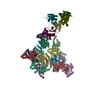
| ||||||||
|---|---|---|---|---|---|---|---|---|---|
| 1 | 
| ||||||||
| 2 | 
| ||||||||
| 3 | 
| ||||||||
| 4 | 
| ||||||||
| Unit cell |
|
-Components
-Transport protein particle ... , 4 types, 20 molecules AGMSBHNTCIOUDEJKPQVW
| #1: Protein | Mass: 24889.262 Da / Num. of mol.: 4 Source method: isolated from a genetically manipulated source Source: (gene. exp.) Gene: TRS23 / Plasmid: pETDuet-1, pCOLADuet-1, pCDFDuet-1 / Production host:  #2: Protein | Mass: 31755.689 Da / Num. of mol.: 4 Source method: isolated from a genetically manipulated source Source: (gene. exp.) Gene: TRS31 / Plasmid: pETDuet-1, pCOLADuet-1, pCDFDuet-1 / Production host: #3: Protein | Mass: 18453.875 Da / Num. of mol.: 4 Source method: isolated from a genetically manipulated source Source: (gene. exp.) Gene: BET5 / Plasmid: pETDuet-1, pCOLADuet-1, pCDFDuet-1 / Production host: #4: Protein | Mass: 22152.445 Da / Num. of mol.: 8 Source method: isolated from a genetically manipulated source Source: (gene. exp.) Gene: BET3 / Plasmid: pETDuet-1, pCOLADuet-1, pCDFDuet-1 / Production host: |
|---|
-Protein / Non-polymers , 2 types, 8 molecules FLRX
| #5: Protein | Mass: 23240.227 Da / Num. of mol.: 4 Source method: isolated from a genetically manipulated source Source: (gene. exp.) Gene: YPT1, YP2 / Plasmid: pETDuet-1, pCOLADuet-1, pCDFDuet-1 / Production host: #6: Chemical | ChemComp-PLM /  Mass: 256.424 Da / Num. of mol.: 4 / Source method: obtained synthetically / Formula: C16H32O2 Mass: 256.424 Da / Num. of mol.: 4 / Source method: obtained synthetically / Formula: C16H32O2 |
|---|
-Details
| Has protein modification | Y |
|---|
-Experimental details
-Experiment
| Experiment | Method: X-RAY DIFFRACTION / Number of used crystals: 1 |
|---|
- Sample preparation
| Crystal | Density Matthews: 3.38 Å3/Da / Density % sol: 63.58 % |
|---|---|
| Crystal grow | Temperature: 295 K / Method: vapor diffusion, hanging drop / pH: 6.5 Details: 3-5% PEG 20,000, 0.1 M MES, 3% (w/v) sorbitol, 5 mM DTT, pH 6.5, VAPOR DIFFUSION, HANGING DROP, temperature 295K |
-Data collection
| Diffraction | Mean temperature: 100 K | |||||||||
|---|---|---|---|---|---|---|---|---|---|---|
| Diffraction source | Source: SYNCHROTRON / Site: APS  / Beamline: 24-ID-C / Wavelength: 0.9795, 0.9537 / Beamline: 24-ID-C / Wavelength: 0.9795, 0.9537 | |||||||||
| Detector | Type: ADSC QUANTUM 315 / Detector: CCD / Date: Jun 7, 2007 | |||||||||
| Radiation | Monochromator: single crystal monochromator / Protocol: MAD / Monochromatic (M) / Laue (L): M / Scattering type: x-ray | |||||||||
| Radiation wavelength |
| |||||||||
| Reflection | Resolution: 3.7→50 Å / Num. all: 81195 / Num. obs: 81195 / % possible obs: 98.5 % / Observed criterion σ(F): 0 / Observed criterion σ(I): -1 / Redundancy: 3.4 % / Rmerge(I) obs: 0.08 / Rsym value: 0.08 / Net I/σ(I): 11.6 | |||||||||
| Reflection shell | Resolution: 3.7→3.83 Å / Redundancy: 2.6 % / Rmerge(I) obs: 0.368 / Mean I/σ(I) obs: 2.2 / Num. unique all: 7251 / Rsym value: 0.368 / % possible all: 91.4 |
- Processing
| Software |
| |||||||||||||||||||||||||
|---|---|---|---|---|---|---|---|---|---|---|---|---|---|---|---|---|---|---|---|---|---|---|---|---|---|---|
| Refinement | Method to determine structure: MAD / Resolution: 3.7→25 Å / σ(F): 0 / Stereochemistry target values: Engh & Huber
| |||||||||||||||||||||||||
| Refinement step | Cycle: LAST / Resolution: 3.7→25 Å
| |||||||||||||||||||||||||
| Refine LS restraints |
| |||||||||||||||||||||||||
| LS refinement shell | Resolution: 3.7→3.72 Å /
|
