+ Open data
- Basic information
| Entry | Database: EMDB / ID: EMD-21900 | |||||||||
|---|---|---|---|---|---|---|---|---|---|---|
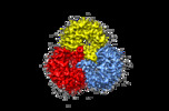
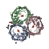
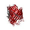
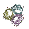
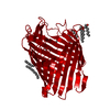
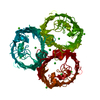
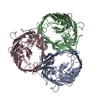
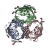
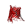
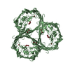
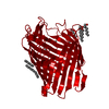
| Title | Cryo-EM structure of E. Coli OmpF | |||||||||
 Map data Map data | E. coli OmpF in nanodisc | |||||||||
 Sample Sample |
| |||||||||
 Keywords Keywords | outer membrane porin / omp / ompf / MEMBRANE PROTEIN | |||||||||
| Function / homology |  Function and homology information Function and homology informationcolicin transmembrane transporter activity / porin activity / monoatomic ion channel complex / pore complex / protein homotrimerization / monoatomic ion channel activity / lipopolysaccharide binding / cell outer membrane / disordered domain specific binding / protein transport ...colicin transmembrane transporter activity / porin activity / monoatomic ion channel complex / pore complex / protein homotrimerization / monoatomic ion channel activity / lipopolysaccharide binding / cell outer membrane / disordered domain specific binding / protein transport / monoatomic ion transmembrane transport / lipid binding / identical protein binding / membrane Similarity search - Function | |||||||||
| Biological species |   | |||||||||
| Method | single particle reconstruction / cryo EM / Resolution: 3.15 Å | |||||||||
 Authors Authors | Morgan CE / Su C-C | |||||||||
| Funding support | 1 items
| |||||||||
 Citation Citation |  Journal: Nat Methods / Year: 2021 Journal: Nat Methods / Year: 2021Title: A 'Build and Retrieve' methodology to simultaneously solve cryo-EM structures of membrane proteins. Authors: Chih-Chia Su / Meinan Lyu / Christopher E Morgan / Jani Reddy Bolla / Carol V Robinson / Edward W Yu /   Abstract: Single-particle cryo-electron microscopy (cryo-EM) has become a powerful technique in the field of structural biology. However, the inability to reliably produce pure, homogeneous membrane protein ...Single-particle cryo-electron microscopy (cryo-EM) has become a powerful technique in the field of structural biology. However, the inability to reliably produce pure, homogeneous membrane protein samples hampers the progress of their structural determination. Here, we develop a bottom-up iterative method, Build and Retrieve (BaR), that enables the identification and determination of cryo-EM structures of a variety of inner and outer membrane proteins, including membrane protein complexes of different sizes and dimensions, from a heterogeneous, impure protein sample. We also use the BaR methodology to elucidate structural information from Escherichia coli K12 crude membrane and raw lysate. The findings demonstrate that it is possible to solve high-resolution structures of a number of relatively small (<100 kDa) and less abundant (<10%) unidentified membrane proteins within a single, heterogeneous sample. Importantly, these results highlight the potential of cryo-EM for systems structural proteomics. | |||||||||
| History |
|
- Structure visualization
| Movie |
 Movie viewer Movie viewer |
|---|---|
| Structure viewer | EM map:  SurfView SurfView Molmil Molmil Jmol/JSmol Jmol/JSmol |
| Supplemental images |
- Downloads & links
-EMDB archive
| Map data |  emd_21900.map.gz emd_21900.map.gz | 2.2 MB |  EMDB map data format EMDB map data format | |
|---|---|---|---|---|
| Header (meta data) |  emd-21900-v30.xml emd-21900-v30.xml emd-21900.xml emd-21900.xml | 9.7 KB 9.7 KB | Display Display |  EMDB header EMDB header |
| Images |  emd_21900.png emd_21900.png | 172.3 KB | ||
| Filedesc metadata |  emd-21900.cif.gz emd-21900.cif.gz | 4.9 KB | ||
| Archive directory |  http://ftp.pdbj.org/pub/emdb/structures/EMD-21900 http://ftp.pdbj.org/pub/emdb/structures/EMD-21900 ftp://ftp.pdbj.org/pub/emdb/structures/EMD-21900 ftp://ftp.pdbj.org/pub/emdb/structures/EMD-21900 | HTTPS FTP |
-Validation report
| Summary document |  emd_21900_validation.pdf.gz emd_21900_validation.pdf.gz | 333.2 KB | Display |  EMDB validaton report EMDB validaton report |
|---|---|---|---|---|
| Full document |  emd_21900_full_validation.pdf.gz emd_21900_full_validation.pdf.gz | 332.8 KB | Display | |
| Data in XML |  emd_21900_validation.xml.gz emd_21900_validation.xml.gz | 6.9 KB | Display | |
| Data in CIF |  emd_21900_validation.cif.gz emd_21900_validation.cif.gz | 7.9 KB | Display | |
| Arichive directory |  https://ftp.pdbj.org/pub/emdb/validation_reports/EMD-21900 https://ftp.pdbj.org/pub/emdb/validation_reports/EMD-21900 ftp://ftp.pdbj.org/pub/emdb/validation_reports/EMD-21900 ftp://ftp.pdbj.org/pub/emdb/validation_reports/EMD-21900 | HTTPS FTP |
-Related structure data
| Related structure data |  6wtzMC  6wtiC  6wu0C  6wu6C  7jz2C  7jz3C  7jz6C  7jzhC M: atomic model generated by this map C: citing same article ( |
|---|---|
| Similar structure data |
- Links
Links
| EMDB pages |  EMDB (EBI/PDBe) / EMDB (EBI/PDBe) /  EMDataResource EMDataResource |
|---|
- Map
Map
| File |  Download / File: emd_21900.map.gz / Format: CCP4 / Size: 163.6 MB / Type: IMAGE STORED AS FLOATING POINT NUMBER (4 BYTES) Download / File: emd_21900.map.gz / Format: CCP4 / Size: 163.6 MB / Type: IMAGE STORED AS FLOATING POINT NUMBER (4 BYTES) | ||||||||||||||||||||||||||||||||||||||||||||||||||||||||||||||||||||
|---|---|---|---|---|---|---|---|---|---|---|---|---|---|---|---|---|---|---|---|---|---|---|---|---|---|---|---|---|---|---|---|---|---|---|---|---|---|---|---|---|---|---|---|---|---|---|---|---|---|---|---|---|---|---|---|---|---|---|---|---|---|---|---|---|---|---|---|---|---|
| Annotation | E. coli OmpF in nanodisc | ||||||||||||||||||||||||||||||||||||||||||||||||||||||||||||||||||||
| Projections & slices | Image control
Images are generated by Spider. | ||||||||||||||||||||||||||||||||||||||||||||||||||||||||||||||||||||
| Voxel size | X=Y=Z: 1.08 Å | ||||||||||||||||||||||||||||||||||||||||||||||||||||||||||||||||||||
| Density |
| ||||||||||||||||||||||||||||||||||||||||||||||||||||||||||||||||||||
| Symmetry | Space group: 1 | ||||||||||||||||||||||||||||||||||||||||||||||||||||||||||||||||||||
| Details | EMDB XML:
CCP4 map header:
| ||||||||||||||||||||||||||||||||||||||||||||||||||||||||||||||||||||
-Supplemental data
- Sample components
-Entire : OmpF
| Entire | Name: OmpF |
|---|---|
| Components |
|
-Supramolecule #1: OmpF
| Supramolecule | Name: OmpF / type: complex / ID: 1 / Parent: 0 / Macromolecule list: #1 |
|---|---|
| Source (natural) | Organism:  |
-Macromolecule #1: Outer membrane porin F
| Macromolecule | Name: Outer membrane porin F / type: protein_or_peptide / ID: 1 / Number of copies: 3 / Enantiomer: LEVO |
|---|---|
| Source (natural) | Organism:  |
| Molecular weight | Theoretical: 39.365043 KDa |
| Sequence | String: MMKRNILAVI VPALLVAGTA NAAEIYNKDG NKVDLYGKAV GLHYFSKGNG ENSYGGNGDM TYARLGFKGE TQINSDLTGY GQWEYNFQG NNSEGADAQT GNKTRLAFAG LKYADVGSFD YGRNYGVVYD ALGYTDMLPE FGGDTAYSDD FFVGRVGGVA T YRNSNFFG ...String: MMKRNILAVI VPALLVAGTA NAAEIYNKDG NKVDLYGKAV GLHYFSKGNG ENSYGGNGDM TYARLGFKGE TQINSDLTGY GQWEYNFQG NNSEGADAQT GNKTRLAFAG LKYADVGSFD YGRNYGVVYD ALGYTDMLPE FGGDTAYSDD FFVGRVGGVA T YRNSNFFG LVDGLNFAVQ YLGKNERDTA RRSNGDGVGG SISYEYEGFG IVGAYGAADR TNLQEAQPLG NGKKAEQWAT GL KYDANNI YLAANYGETR NATPITNKFT NTSGFANKTQ DVLLVAQYQF DFGLRPSIAY TKSKAKDVEG IGDVDLVNYF EVG ATYYFN KNMSTYVDYI INQIDSDNKL GVGSDDTVAV GIVYQF UniProtKB: Outer membrane porin F |
-Macromolecule #2: water
| Macromolecule | Name: water / type: ligand / ID: 2 / Number of copies: 36 / Formula: HOH |
|---|---|
| Molecular weight | Theoretical: 18.015 Da |
| Chemical component information |  ChemComp-HOH: |
-Experimental details
-Structure determination
| Method | cryo EM |
|---|---|
 Processing Processing | single particle reconstruction |
| Aggregation state | particle |
- Sample preparation
| Buffer | pH: 7.5 |
|---|---|
| Vitrification | Cryogen name: ETHANE |
- Electron microscopy
| Microscope | TFS KRIOS |
|---|---|
| Image recording | Film or detector model: GATAN K3 (6k x 4k) / Average electron dose: 50.0 e/Å2 |
| Electron beam | Acceleration voltage: 300 kV / Electron source:  FIELD EMISSION GUN FIELD EMISSION GUN |
| Electron optics | Illumination mode: SPOT SCAN / Imaging mode: BRIGHT FIELD |
| Experimental equipment |  Model: Titan Krios / Image courtesy: FEI Company |
- Image processing
| Startup model | Type of model: NONE |
|---|---|
| Final reconstruction | Applied symmetry - Point group: C3 (3 fold cyclic) / Resolution.type: BY AUTHOR / Resolution: 3.15 Å / Resolution method: FSC 0.143 CUT-OFF / Software - Name: cryoSPARC / Number images used: 43793 |
| Initial angle assignment | Type: MAXIMUM LIKELIHOOD / Software - Name: cryoSPARC |
| Final angle assignment | Type: MAXIMUM LIKELIHOOD / Software - Name: cryoSPARC |
 Movie
Movie Controller




 X (Sec.)
 Y (Row.)
 Z (Col.)


