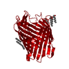
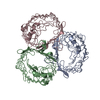
MEMBRANE PROTEIN / OmpF PORIN / Ordered Waters / Ion transport / Membrane / Outer membrane / Phage recognition / Transmembrane / Transport
Function / homology
Function and homology information
colicin transmembrane transporter activity / porin activity / protein homotrimerization / pore complex / monoatomic ion channel complex / monoatomic ion channel activity / cell outer membrane / lipopolysaccharide binding / disordered domain specific binding / protein transport ...colicin transmembrane transporter activity / porin activity / protein homotrimerization / pore complex / monoatomic ion channel complex / monoatomic ion channel activity / cell outer membrane / lipopolysaccharide binding / disordered domain specific binding / protein transport / monoatomic ion transmembrane transport / lipid binding / membrane / identical protein binding Similarity search - Function
Porin, gammaproteobacterial / Porin, Gram-negative type, conserved site / General diffusion Gram-negative porins signature. / Porin domain, Gram-negative type / Gram-negative porin / Porin / Porin, Gram-negative type / : / Porin domain superfamily / Porin ...Porin, gammaproteobacterial / Porin, Gram-negative type, conserved site / General diffusion Gram-negative porins signature. / Porin domain, Gram-negative type / Gram-negative porin / Porin / Porin, Gram-negative type / : / Porin domain superfamily / Porin / Beta Barrel / Mainly Beta Similarity search - Domain/homology
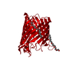
Resolution: 1.59→36.1 Å / Cor.coef. Fo:Fc: 0.966 / Cor.coef. Fo:Fc free: 0.948 / SU B: 1.36 / SU ML: 0.05 / Cross valid method: THROUGHOUT / ESU R: 0.081 / ESU R Free: 0.086 / Stereochemistry target values: MAXIMUM LIKELIHOOD Details: THE ELECTRON DENSITY AT RESIDUE ASN27 IS SMALL; THE OCCUPANCY AT THIS SITE IN OTHER OMPF STRUCTURES, 1HXX AND 2OMF, IS ZERO. HYDROGENS HAVE BEEN ADDED IN THE RIDING POSITIONS.
Rfactor
Num. reflection
% reflection
Selection details
Rfree
0.21837
2740
5.1 %
RANDOM
Rwork
0.18078
-
-
-
obs
0.18256
51108
99.34 %
-
Solvent computation
Ion probe radii: 0.8 Å / Shrinkage radii: 0.8 Å / VDW probe radii: 1.2 Å / Solvent model: MASK
Displacement parameters
Biso mean: 27.534 Å2
Baniso -1
Baniso -2
Baniso -3
1-
-1.1 Å2
-0.55 Å2
0 Å2
2-
-
-1.1 Å2
0 Å2
3-
-
-
1.66 Å2
Refinement step
Cycle: LAST / Resolution: 1.59→36.1 Å
Protein
Nucleic acid
Ligand
Solvent
Total
Num. atoms
2641
0
43
322
3006
Refine LS restraints
Refine-ID
Type
Dev ideal
Dev ideal target
Number
X-RAY DIFFRACTION
r_bond_refined_d
0.01
0.022
2736
X-RAY DIFFRACTION
r_bond_other_d
X-RAY DIFFRACTION
r_angle_refined_deg
1.218
1.943
3689
X-RAY DIFFRACTION
r_angle_other_deg
X-RAY DIFFRACTION
r_dihedral_angle_1_deg
5.899
5
337
X-RAY DIFFRACTION
r_dihedral_angle_2_deg
37.233
25
146
X-RAY DIFFRACTION
r_dihedral_angle_3_deg
13.074
15
407
X-RAY DIFFRACTION
r_dihedral_angle_4_deg
15.258
15
11
X-RAY DIFFRACTION
r_chiral_restr
0.09
0.2
379
X-RAY DIFFRACTION
r_gen_planes_refined
0.005
0.02
2150
X-RAY DIFFRACTION
r_gen_planes_other
X-RAY DIFFRACTION
r_nbd_refined
0.193
0.2
1114
X-RAY DIFFRACTION
r_nbd_other
X-RAY DIFFRACTION
r_nbtor_refined
0.305
0.2
1893
X-RAY DIFFRACTION
r_nbtor_other
X-RAY DIFFRACTION
r_xyhbond_nbd_refined
0.115
0.2
273
X-RAY DIFFRACTION
r_xyhbond_nbd_other
X-RAY DIFFRACTION
r_metal_ion_refined
0.082
0.2
2
X-RAY DIFFRACTION
r_metal_ion_other
X-RAY DIFFRACTION
r_symmetry_vdw_refined
0.146
0.2
73
X-RAY DIFFRACTION
r_symmetry_vdw_other
X-RAY DIFFRACTION
r_symmetry_hbond_refined
0.143
0.2
18
X-RAY DIFFRACTION
r_symmetry_hbond_other
X-RAY DIFFRACTION
r_symmetry_metal_ion_refined
X-RAY DIFFRACTION
r_symmetry_metal_ion_other
X-RAY DIFFRACTION
r_mcbond_it
0.807
1.5
1684
X-RAY DIFFRACTION
r_mcbond_other
X-RAY DIFFRACTION
r_mcangle_it
1.378
2
2628
X-RAY DIFFRACTION
r_scbond_it
2.162
3
1200
X-RAY DIFFRACTION
r_scangle_it
3.092
4.5
1061
X-RAY DIFFRACTION
r_rigid_bond_restr
X-RAY DIFFRACTION
r_sphericity_free
X-RAY DIFFRACTION
r_sphericity_bonded
LS refinement shell
Resolution: 1.592→1.634 Å / Total num. of bins used: 20
Rfactor
Num. reflection
% reflection
Rfree
0.261
179
-
Rwork
0.227
3547
-
obs
-
-
93.64 %
+
About Yorodumi
-
News
-
Feb 9, 2022. New format data for meta-information of EMDB entries
New format data for meta-information of EMDB entries
Version 3 of the EMDB header file is now the official format.
The previous official version 1.9 will be removed from the archive.
In the structure databanks used in Yorodumi, some data are registered as the other names, "COVID-19 virus" and "2019-nCoV". Here are the details of the virus and the list of structure data.
Jan 31, 2019. EMDB accession codes are about to change! (news from PDBe EMDB page)
EMDB accession codes are about to change! (news from PDBe EMDB page)
The allocation of 4 digits for EMDB accession codes will soon come to an end. Whilst these codes will remain in use, new EMDB accession codes will include an additional digit and will expand incrementally as the available range of codes is exhausted. The current 4-digit format prefixed with “EMD-” (i.e. EMD-XXXX) will advance to a 5-digit format (i.e. EMD-XXXXX), and so on. It is currently estimated that the 4-digit codes will be depleted around Spring 2019, at which point the 5-digit format will come into force.
The EM Navigator/Yorodumi systems omit the EMD- prefix.
Related info.:Q: What is EMD? / ID/Accession-code notation in Yorodumi/EM Navigator
Yorodumi is a browser for structure data from EMDB, PDB, SASBDB, etc.
This page is also the successor to EM Navigator detail page, and also detail information page/front-end page for Omokage search.
The word "yorodu" (or yorozu) is an old Japanese word meaning "ten thousand". "mi" (miru) is to see.
Related info.:EMDB / PDB / SASBDB / Comparison of 3 databanks / Yorodumi Search / Aug 31, 2016. New EM Navigator & Yorodumi / Yorodumi Papers / Jmol/JSmol / Function and homology information / Changes in new EM Navigator and Yorodumi
 Movie
Movie Controller


 Open data
 Basic information
 Components
 Keywords
 Function and homology information
 X-RAY DIFFRACTION /
  Authors
 Citation
 Structure visualization
 Downloads & links
 Other downloads


 PDBj
PDBj Assembly
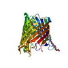

 Mass: 24.305 Da / Num. of mol.: 1 / Source method: obtained synthetically / Formula: Mg
 Mass: 306.438 Da / Num. of mol.: 2 / Source method: obtained synthetically / Formula: C16H34O5 / Comment: C8E, detergent*YM
 Mass: 18.015 Da / Num. of mol.: 322 / Source method: isolated from a natural source / Formula: H2O
 Sample preparation
 / Beamline: 19-ID / Wavelength: 0.97856 Å
 Processing