 Movie
Movie Controller


+ Open data

- Basic information
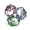
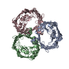
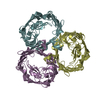
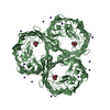
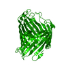
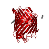
| Entry | Database: PDB / ID: 2zld | ||||||
|---|---|---|---|---|---|---|---|
| Title | Structure of OmpF co-crystallized with T83 | ||||||
 Components Components |
| ||||||
 Keywords Keywords | MEMBRANE PROTEIN / BtuB / colicin E3 / ribosomal RNAase / disordered T83 / Tol system / Ion transport / Membrane / Outer membrane / Phage recognition / Porin / Transmembrane / Transport | ||||||
| Function / homology |  Function and homology information Function and homology informationcolicin transmembrane transporter activity / porin activity / monoatomic ion channel complex / protein homotrimerization / pore complex / monoatomic ion channel activity / cell outer membrane / lipopolysaccharide binding / disordered domain specific binding / protein transport ...colicin transmembrane transporter activity / porin activity / monoatomic ion channel complex / protein homotrimerization / pore complex / monoatomic ion channel activity / cell outer membrane / lipopolysaccharide binding / disordered domain specific binding / protein transport / monoatomic ion transmembrane transport / lipid binding / membrane / identical protein binding Similarity search - Function | ||||||
| Biological species |  | ||||||
| Method |  X-RAY DIFFRACTION / SYNCHROTRON / MOLECULAR REPLACEMENT / Resolution: 3 Å X-RAY DIFFRACTION / SYNCHROTRON / MOLECULAR REPLACEMENT / Resolution: 3 Å | ||||||
 Authors Authors | Cramer, W.A. / Zakharov, S.D. / Yamashita, E. | ||||||
 Citation Citation | Journal: Embo J. / Year: 2008 Title: Crystal structures of the OmpF porin: function in a colicin translocon. Authors: Yamashita, E. / Zhalnina, M.V. / Zakharov, S.D. / Sharma, O. / Cramer, W.A. | ||||||
| History |
|
- Structure visualization
| Structure viewer | Molecule: MolmilJmol/JSmol |
|---|
- Downloads & links
-Download
| PDBx/mmCIF format | 2zld.cif.gz | 137 KB | Display | PDBx/mmCIF format |
|---|---|---|---|---|
| PDB format | pdb2zld.ent.gz | 108.7 KB | Display | PDB format |
| PDBx/mmJSON format | 2zld.json.gz | Tree view | PDBx/mmJSON format | |
| Others |  Other downloads Other downloads |
-Validation report
| Arichive directory | https://data.pdbj.org/pub/pdb/validation_reports/zl/2zldftp://data.pdbj.org/pub/pdb/validation_reports/zl/2zld | HTTPS FTP |
|---|
-Related structure data
| Related structure data |  2zfgC  2omfS C: citing same article ( S: Starting model for refinement |
|---|---|
| Similar structure data |
-Links
 PDBj
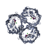
PDBj- Assembly
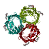
| Deposited unit | 
| ||||||||||||||||||
|---|---|---|---|---|---|---|---|---|---|---|---|---|---|---|---|---|---|---|---|
| 1 | 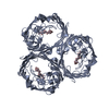
| ||||||||||||||||||
| 2 | 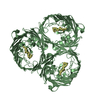
| ||||||||||||||||||
| 3 |
| ||||||||||||||||||
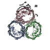
| Unit cell |
| ||||||||||||||||||
| Noncrystallographic symmetry (NCS) | NCS domain:
NCS domain segments: Component-ID: 1 / Ens-ID: 1 / Beg auth comp-ID: ALA / Beg label comp-ID: ALA / End auth comp-ID: PHE / End label comp-ID: PHE / Refine code: 1 / Auth seq-ID: 1 - 340 / Label seq-ID: 1 - 340
|
-Components
| #1: Protein | Mass: 37114.250 Da / Num. of mol.: 2 Source method: isolated from a genetically manipulated source Source: (gene. exp.) #2: Protein/peptide | Mass: 613.749 Da / Num. of mol.: 2 / Fragment: T83 (RESIDUES 1-83) Source method: isolated from a genetically manipulated source Source: (gene. exp.) Sequence details | THE AUTHOR CO-CRYSTALLIZED THE OMPF WITH THE N-TERMINAL TRANSLOCATION DOMAIN OF COLICIN E3 (T83). ...THE AUTHOR CO-CRYSTALLIZ | |
|---|
-Experimental details
-Experiment
| Experiment | Method: X-RAY DIFFRACTION / Number of used crystals: 1 |
|---|
- Sample preparation
| Crystal | Density Matthews: 2.99 Å3/Da / Density % sol: 58.82 % |
|---|
-Data collection
| Diffraction | Mean temperature: 100 K |
|---|---|
| Diffraction source | Source: SYNCHROTRON / Site: APS  / Beamline: 19-ID / Wavelength: 0.97935 Å / Beamline: 19-ID / Wavelength: 0.97935 Å |
| Detector | Type: ADSC QUANTUM 315 / Detector: CCD / Date: Nov 25, 2007 |
| Radiation | Protocol: SINGLE WAVELENGTH / Monochromatic (M) / Laue (L): M / Scattering type: x-ray |
| Radiation wavelength | Wavelength: 0.97935 Å / Relative weight: 1 |
| Reflection | Highest resolution: 3 Å / Num. obs: 17885 / % possible obs: 99.9 % / Observed criterion σ(I): -3 / Redundancy: 8.3 % / Rmerge(I) obs: 0.076 |
- Processing
| Software |
| ||||||||||||||||||||||||||||||||||||||||||||||||||||||||||||||||||||||||||||||||||||||||||||||||||||||||||||||||||||||||||||||||||||||||||||||||||||||||||||||||||||||||||
|---|---|---|---|---|---|---|---|---|---|---|---|---|---|---|---|---|---|---|---|---|---|---|---|---|---|---|---|---|---|---|---|---|---|---|---|---|---|---|---|---|---|---|---|---|---|---|---|---|---|---|---|---|---|---|---|---|---|---|---|---|---|---|---|---|---|---|---|---|---|---|---|---|---|---|---|---|---|---|---|---|---|---|---|---|---|---|---|---|---|---|---|---|---|---|---|---|---|---|---|---|---|---|---|---|---|---|---|---|---|---|---|---|---|---|---|---|---|---|---|---|---|---|---|---|---|---|---|---|---|---|---|---|---|---|---|---|---|---|---|---|---|---|---|---|---|---|---|---|---|---|---|---|---|---|---|---|---|---|---|---|---|---|---|---|---|---|---|---|---|---|---|
| Refinement | Method to determine structure: MOLECULAR REPLACEMENT Starting model: 2OMF Resolution: 3→101.02 Å / Cor.coef. Fo:Fc: 0.868 / Cor.coef. Fo:Fc free: 0.857 / SU B: 23.547 / SU ML: 0.438 / Cross valid method: THROUGHOUT / ESU R Free: 0.502 / Stereochemistry target values: MAXIMUM LIKELIHOOD / Details: HYDROGENS HAVE BEEN ADDED IN THE RIDING POSITIONS
| ||||||||||||||||||||||||||||||||||||||||||||||||||||||||||||||||||||||||||||||||||||||||||||||||||||||||||||||||||||||||||||||||||||||||||||||||||||||||||||||||||||||||||
| Solvent computation | Ion probe radii: 0.8 Å / Shrinkage radii: 0.8 Å / VDW probe radii: 1.4 Å / Solvent model: MASK | ||||||||||||||||||||||||||||||||||||||||||||||||||||||||||||||||||||||||||||||||||||||||||||||||||||||||||||||||||||||||||||||||||||||||||||||||||||||||||||||||||||||||||
| Displacement parameters | Biso mean: 48.67 Å2
| ||||||||||||||||||||||||||||||||||||||||||||||||||||||||||||||||||||||||||||||||||||||||||||||||||||||||||||||||||||||||||||||||||||||||||||||||||||||||||||||||||||||||||
| Refinement step | Cycle: LAST / Resolution: 3→101.02 Å
| ||||||||||||||||||||||||||||||||||||||||||||||||||||||||||||||||||||||||||||||||||||||||||||||||||||||||||||||||||||||||||||||||||||||||||||||||||||||||||||||||||||||||||
| Refine LS restraints |
| ||||||||||||||||||||||||||||||||||||||||||||||||||||||||||||||||||||||||||||||||||||||||||||||||||||||||||||||||||||||||||||||||||||||||||||||||||||||||||||||||||||||||||
| Refine LS restraints NCS | Dom-ID: 1 / Auth asym-ID: A / Ens-ID: 1 / Number: 2619 / Refine-ID: X-RAY DIFFRACTION
| ||||||||||||||||||||||||||||||||||||||||||||||||||||||||||||||||||||||||||||||||||||||||||||||||||||||||||||||||||||||||||||||||||||||||||||||||||||||||||||||||||||||||||
| LS refinement shell | Resolution: 3→3.078 Å / Total num. of bins used: 20
|
