 Movie
Movie Controller


+ Open data

- Basic information
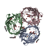
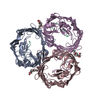
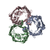
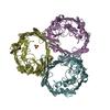
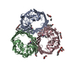
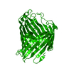
| Entry | Database: PDB / ID: 1hxx | ||||||
|---|---|---|---|---|---|---|---|
| Title | OMPF PORIN MUTANT Y106F | ||||||
 Components Components | OUTER MEMBRANE PROTEIN F | ||||||
 Keywords Keywords | MEMBRANE PROTEIN / porin / beta barrel | ||||||
| Function / homology |  Function and homology information Function and homology informationcolicin transmembrane transporter activity / porin activity / monoatomic ion channel complex / protein homotrimerization / pore complex / monoatomic ion channel activity / cell outer membrane / lipopolysaccharide binding / disordered domain specific binding / protein transport ...colicin transmembrane transporter activity / porin activity / monoatomic ion channel complex / protein homotrimerization / pore complex / monoatomic ion channel activity / cell outer membrane / lipopolysaccharide binding / disordered domain specific binding / protein transport / monoatomic ion transmembrane transport / lipid binding / membrane / identical protein binding Similarity search - Function | ||||||
| Biological species |  | ||||||
| Method |  X-RAY DIFFRACTION / SYNCHROTRON / difference fourier / Resolution: 2.2 Å X-RAY DIFFRACTION / SYNCHROTRON / difference fourier / Resolution: 2.2 Å | ||||||
 Authors Authors | Phale, P.S. / Philippsen, A. / Widmer, C. / Phale, V.P. / Rosenbusch, J.P. / Schirmer, T. | ||||||
 Citation Citation | Journal: Biochemistry / Year: 2001 Title: Role of charged residues at the OmpF porin channel constriction probed by mutagenesis and simulation. Authors: Phale, P.S. / Philippsen, A. / Widmer, C. / Phale, V.P. / Rosenbusch, J.P. / Schirmer, T. | ||||||
| History |
|
- Structure visualization
| Structure viewer | Molecule: MolmilJmol/JSmol |
|---|
- Downloads & links
-Download
| PDBx/mmCIF format | 1hxx.cif.gz | 82.2 KB | Display | PDBx/mmCIF format |
|---|---|---|---|---|
| PDB format | pdb1hxx.ent.gz | 61.2 KB | Display | PDB format |
| PDBx/mmJSON format | 1hxx.json.gz | Tree view | PDBx/mmJSON format | |
| Others |  Other downloads Other downloads |
-Validation report
| Arichive directory | https://data.pdbj.org/pub/pdb/validation_reports/hx/1hxxftp://data.pdbj.org/pub/pdb/validation_reports/hx/1hxx | HTTPS FTP |
|---|
-Related structure data


-Links
 PDBj
PDBj- Assembly
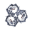
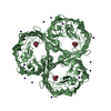
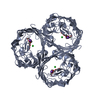
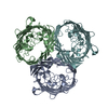
| Deposited unit | 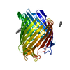
| ||||||||
|---|---|---|---|---|---|---|---|---|---|
| 1 | 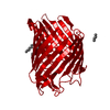
| ||||||||
| Unit cell |
|
-Components
| #1: Protein | Mass: 37098.250 Da / Num. of mol.: 1 / Mutation: Y106F Source method: isolated from a genetically manipulated source Source: (gene. exp.) | ||
|---|---|---|---|
| #2: Chemical | ChemComp-C8E / (   Mass: 306.438 Da / Num. of mol.: 4 / Source method: obtained synthetically / Formula: C16H34O5 / Comment: C8E, detergent*YM Mass: 306.438 Da / Num. of mol.: 4 / Source method: obtained synthetically / Formula: C16H34O5 / Comment: C8E, detergent*YM#3: Water | ChemComp-HOH / |  Mass: 18.015 Da / Num. of mol.: 145 / Source method: isolated from a natural source / Formula: H2O Mass: 18.015 Da / Num. of mol.: 145 / Source method: isolated from a natural source / Formula: H2O |
-Experimental details
-Experiment
| Experiment | Method: X-RAY DIFFRACTION / Number of used crystals: 1 |
|---|
- Sample preparation
| Crystal | Density Matthews: 2.85 Å3/Da / Density % sol: 56.88 % | |||||||||||||||
|---|---|---|---|---|---|---|---|---|---|---|---|---|---|---|---|---|
| Crystal grow | *PLUS Method: microdialysis / Details: Pauptit, R.A., (1991) J. Mol. Biol., 218, 505. | |||||||||||||||
| Components of the solutions | *PLUS
|
-Data collection
| Diffraction | Mean temperature: 100 K |
|---|---|
| Diffraction source | Source: SYNCHROTRON / Site: EMBL/DESY, HAMBURG  / Beamline: X11 / Wavelength: 1.042 Å / Beamline: X11 / Wavelength: 1.042 Å |
| Detector | Type: MACSCIENCE / Detector: IMAGE PLATE / Date: Jan 1, 1998 |
| Radiation | Protocol: SINGLE WAVELENGTH / Monochromatic (M) / Laue (L): M / Scattering type: x-ray |
| Radiation wavelength | Wavelength: 1.042 Å / Relative weight: 1 |
| Reflection | Resolution: 2.2→30 Å / Num. all: 71614 / Num. obs: 19807 / % possible obs: 79.9 % / Observed criterion σ(F): 6 / Observed criterion σ(I): 0 / Redundancy: 3.4 % / Biso Wilson estimate: 28 Å2 / Rmerge(I) obs: 0.065 / Net I/σ(I): 7 |
| Reflection | *PLUS % possible obs: 97.9 % / Num. measured all: 71614 |
- Processing
| Software |
| ||||||||||||||||||||
|---|---|---|---|---|---|---|---|---|---|---|---|---|---|---|---|---|---|---|---|---|---|
| Refinement | Method to determine structure: difference fourier / Resolution: 2.2→8 Å / Stereochemistry target values: engh & huber
| ||||||||||||||||||||
| Refinement step | Cycle: LAST / Resolution: 2.2→8 Å
| ||||||||||||||||||||
| Refine LS restraints |
| ||||||||||||||||||||
| Software | *PLUS Name: REFMAC / Classification: refinement | ||||||||||||||||||||
| Refinement | *PLUS Rfactor obs: 0.174 / Rfactor Rfree: 0.244 | ||||||||||||||||||||
| Solvent computation | *PLUS | ||||||||||||||||||||
| Displacement parameters | *PLUS | ||||||||||||||||||||
| Refine LS restraints | *PLUS Type: p_bond_d / Dev ideal: 0.08 |
