 Movie
Movie Controller


+ Open data

- Basic information
| Entry | Database: PDB / ID: 4lsi | ||||||
|---|---|---|---|---|---|---|---|
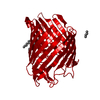
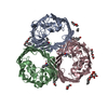
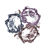
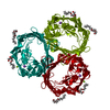
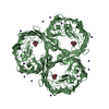
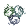
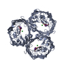
| Title | Ion selectivity of OmpF porin soaked in 0.3M KBr | ||||||
 Components Components | Outer membrane protein F | ||||||
 Keywords Keywords | TRANSPORT PROTEIN / porin / outer membrane protein / beta-barrel / ion transport | ||||||
| Function / homology |  Function and homology information Function and homology informationcolicin transmembrane transporter activity / porin activity / protein homotrimerization / pore complex / monoatomic ion channel complex / monoatomic ion channel activity / cell outer membrane / lipopolysaccharide binding / disordered domain specific binding / protein transport ...colicin transmembrane transporter activity / porin activity / protein homotrimerization / pore complex / monoatomic ion channel complex / monoatomic ion channel activity / cell outer membrane / lipopolysaccharide binding / disordered domain specific binding / protein transport / monoatomic ion transmembrane transport / lipid binding / membrane / identical protein binding Similarity search - Function | ||||||
| Biological species |  | ||||||
| Method |  X-RAY DIFFRACTION / SYNCHROTRON / MOLECULAR REPLACEMENT / Resolution: 2.089 Å X-RAY DIFFRACTION / SYNCHROTRON / MOLECULAR REPLACEMENT / Resolution: 2.089 Å | ||||||
 Authors Authors | Balasundaresan, D. / Blachowicz, L. / Roux, B. | ||||||
 Citation Citation | Journal: J.Am.Chem.Soc. / Year: 2013 Title: A structural study of ion permeation in OmpF porin from anomalous X-ray diffraction and molecular dynamics simulations. Authors: Dhakshnamoorthy, B. / Ziervogel, B.K. / Blachowicz, L. / Roux, B. | ||||||
| History |
|
- Structure visualization
| Structure viewer | Molecule: MolmilJmol/JSmol |
|---|
- Downloads & links
-Download
| PDBx/mmCIF format | 4lsi.cif.gz | 220.9 KB | Display | PDBx/mmCIF format |
|---|---|---|---|---|
| PDB format | pdb4lsi.ent.gz | 175.5 KB | Display | PDB format |
| PDBx/mmJSON format | 4lsi.json.gz | Tree view | PDBx/mmJSON format | |
| Others |  Other downloads Other downloads |
-Validation report
| Arichive directory | https://data.pdbj.org/pub/pdb/validation_reports/ls/4lsiftp://data.pdbj.org/pub/pdb/validation_reports/ls/4lsi | HTTPS FTP |
|---|
-Related structure data
| Related structure data |  4lseC 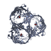 4lsfC 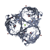 4lshC  2omfS C: citing same article ( S: Starting model for refinement |
|---|---|
| Similar structure data |


-Links
 PDBj
PDBj

- Assembly
| Deposited unit | 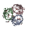
| ||||||||
|---|---|---|---|---|---|---|---|---|---|
| 1 |
| ||||||||
| Unit cell |
| ||||||||
| Components on special symmetry positions |
|
-Components
-Protein , 1 types, 3 molecules ABC
| #1: Protein | Mass: 37171.301 Da / Num. of mol.: 3 / Fragment: UNP residues 23-362 Source method: isolated from a genetically manipulated source Source: (gene. exp.) |
|---|
-Non-polymers , 6 types, 607 molecules 


| #2: Chemical | ChemComp-PEG /  Mass: 106.120 Da / Num. of mol.: 9 / Source method: obtained synthetically / Formula: C4H10O3 Mass: 106.120 Da / Num. of mol.: 9 / Source method: obtained synthetically / Formula: C4H10O3#3: Chemical | ChemComp-C8E / (  Mass: 306.438 Da / Num. of mol.: 4 / Source method: obtained synthetically / Formula: C16H34O5 / Comment: C8E, detergent*YM Mass: 306.438 Da / Num. of mol.: 4 / Source method: obtained synthetically / Formula: C16H34O5 / Comment: C8E, detergent*YM#4: Chemical | ChemComp-GOL /  Mass: 92.094 Da / Num. of mol.: 4 / Source method: obtained synthetically / Formula: C3H8O3 Mass: 92.094 Da / Num. of mol.: 4 / Source method: obtained synthetically / Formula: C3H8O3#5: Chemical | ChemComp-MG /  Mass: 24.305 Da / Num. of mol.: 8 / Source method: obtained synthetically / Formula: Mg Mass: 24.305 Da / Num. of mol.: 8 / Source method: obtained synthetically / Formula: Mg#6: Chemical | ChemComp-BR /  Mass: 79.904 Da / Num. of mol.: 9 / Source method: obtained synthetically / Formula: Br Mass: 79.904 Da / Num. of mol.: 9 / Source method: obtained synthetically / Formula: Br#7: Water | ChemComp-HOH / | Mass: 18.015 Da / Num. of mol.: 573 / Source method: isolated from a natural source / Formula: H2O |
|---|
-Experimental details
-Experiment
| Experiment | Method: X-RAY DIFFRACTION / Number of used crystals: 1 |
|---|
- Sample preparation
| Crystal | Density Matthews: 2.81 Å3/Da / Density % sol: 56.21 % |
|---|---|
| Crystal grow | Temperature: 293 K / Method: vapor diffusion, hanging drop / pH: 6.5 Details: 50% PEG200, 0.1 M sodium cacodylate, 0.2 M magnesium chloride, pH 6.5, crystals soaked in 0.3 M potassium bromide, VAPOR DIFFUSION, HANGING DROP, temperature 293.0K |
-Data collection
| Diffraction | Mean temperature: 100 K |
|---|---|
| Diffraction source | Source: SYNCHROTRON / Site: APS  / Beamline: 24-ID-C / Wavelength: 0.91956 Å / Beamline: 24-ID-C / Wavelength: 0.91956 Å |
| Detector | Type: ADSC QUANTUM 315 / Detector: CCD / Date: Jun 20, 2011 |
| Radiation | Monochromator: double crystal Si(111) / Protocol: SINGLE WAVELENGTH / Monochromatic (M) / Laue (L): M / Scattering type: x-ray |
| Radiation wavelength | Wavelength: 0.91956 Å / Relative weight: 1 |
| Reflection | Resolution: 2.089→50 Å / Num. obs: 71693 / % possible obs: 98.7 % / Observed criterion σ(F): 1 / Observed criterion σ(I): 1 / Redundancy: 3.1 % / Rmerge(I) obs: 0.117 / Net I/σ(I): 13.6 |
| Reflection shell | Resolution: 2.089→2.14 Å / Redundancy: 3.1 % / Rmerge(I) obs: 0.654 / Mean I/σ(I) obs: 1.78 / % possible all: 99.6 |
- Processing
| Software |
| ||||||||||||||||||||||||||||||||||||||||||||||||||||||||||||
|---|---|---|---|---|---|---|---|---|---|---|---|---|---|---|---|---|---|---|---|---|---|---|---|---|---|---|---|---|---|---|---|---|---|---|---|---|---|---|---|---|---|---|---|---|---|---|---|---|---|---|---|---|---|---|---|---|---|---|---|---|---|
| Refinement | Method to determine structure: MOLECULAR REPLACEMENT Starting model: PDB ENTRY 2OMF Resolution: 2.089→32.74 Å / Cor.coef. Fo:Fc: 0.954 / Cor.coef. Fo:Fc free: 0.925 / SU B: 4.708 / SU ML: 0.124 / Cross valid method: THROUGHOUT / σ(F): 2 / ESU R: 0.202 / ESU R Free: 0.185 / Stereochemistry target values: MAXIMUM LIKELIHOOD / Details: HYDROGENS HAVE BEEN ADDED IN THE RIDING POSITIONS
| ||||||||||||||||||||||||||||||||||||||||||||||||||||||||||||
| Solvent computation | Ion probe radii: 0.8 Å / Shrinkage radii: 0.8 Å / VDW probe radii: 1.2 Å / Solvent model: MASK | ||||||||||||||||||||||||||||||||||||||||||||||||||||||||||||
| Displacement parameters | Biso mean: 34.356 Å2
| ||||||||||||||||||||||||||||||||||||||||||||||||||||||||||||
| Refinement step | Cycle: LAST / Resolution: 2.089→32.74 Å
| ||||||||||||||||||||||||||||||||||||||||||||||||||||||||||||
| Refine LS restraints |
| ||||||||||||||||||||||||||||||||||||||||||||||||||||||||||||
| LS refinement shell | Resolution: 2.089→2.143 Å / Total num. of bins used: 20
|
