 Movie
Movie Controller

[English] 日本語
 Yorodumi
- PDB-3hwb: Cation selective pathway of OmpF porin revealed by anomalous diff... -
+ Open data

- Basic information
| Entry | Database: PDB / ID: 3hwb | ||||||
|---|---|---|---|---|---|---|---|
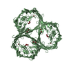
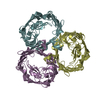
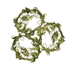
| Title | Cation selective pathway of OmpF porin revealed by anomalous diffraction | ||||||
 Components Components | Outer membrane protein F | ||||||
 Keywords Keywords | MEMBRANE PROTEIN / ion transport / porin / integral membrane protein porin / Cell membrane / Cell outer membrane / Membrane / Phage recognition / Transmembrane / Transport | ||||||
| Function / homology |  Function and homology information Function and homology informationcolicin transmembrane transporter activity / porin activity / protein homotrimerization / pore complex / monoatomic ion channel complex / monoatomic ion channel activity / cell outer membrane / lipopolysaccharide binding / disordered domain specific binding / protein transport ...colicin transmembrane transporter activity / porin activity / protein homotrimerization / pore complex / monoatomic ion channel complex / monoatomic ion channel activity / cell outer membrane / lipopolysaccharide binding / disordered domain specific binding / protein transport / monoatomic ion transmembrane transport / lipid binding / membrane / identical protein binding Similarity search - Function | ||||||
| Biological species |  | ||||||
| Method |  X-RAY DIFFRACTION / SYNCHROTRON / SAD / Resolution: 3 Å X-RAY DIFFRACTION / SYNCHROTRON / SAD / Resolution: 3 Å | ||||||
 Authors Authors | Balasundaresan, D. / Raychaudhury, S. / Blachowicz, L. / Roux, B. | ||||||
 Citation Citation | Journal: J.Mol.Biol. / Year: 2010 Title: Cation-selective pathway of OmpF porin revealed by anomalous X-ray diffraction. Authors: Dhakshnamoorthy, B. / Raychaudhury, S. / Blachowicz, L. / Roux, B. | ||||||
| History |
|
- Structure visualization
| Structure viewer | Molecule: MolmilJmol/JSmol |
|---|
- Downloads & links
-Download
| PDBx/mmCIF format | 3hwb.cif.gz | 142.7 KB | Display | PDBx/mmCIF format |
|---|---|---|---|---|
| PDB format | pdb3hwb.ent.gz | 111.3 KB | Display | PDB format |
| PDBx/mmJSON format | 3hwb.json.gz | Tree view | PDBx/mmJSON format | |
| Others |  Other downloads Other downloads |
-Validation report
| Arichive directory | https://data.pdbj.org/pub/pdb/validation_reports/hw/3hwbftp://data.pdbj.org/pub/pdb/validation_reports/hw/3hwb | HTTPS FTP |
|---|
-Related structure data
| Related structure data |  3hw9C  2omfS C: citing same article ( S: Starting model for refinement |
|---|---|
| Similar structure data |


-Links
 PDBj
PDBj
- Assembly
| Deposited unit | 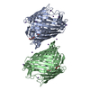
| ||||||||
|---|---|---|---|---|---|---|---|---|---|
| 1 | 
| ||||||||
| 2 | 
| ||||||||
| 3 |
| ||||||||
| Unit cell |
|
-Components
| #1: Protein | Mass: 39365.043 Da / Num. of mol.: 2 Source method: isolated from a genetically manipulated source Source: (gene. exp.) #2: Chemical | ChemComp-RB /   Mass: 85.468 Da / Num. of mol.: 19 / Source method: obtained synthetically / Formula: Rb Mass: 85.468 Da / Num. of mol.: 19 / Source method: obtained synthetically / Formula: Rb#3: Chemical |   Mass: 282.331 Da / Num. of mol.: 2 / Source method: obtained synthetically / Formula: C12H26O7 / Comment: precipitant*YM Mass: 282.331 Da / Num. of mol.: 2 / Source method: obtained synthetically / Formula: C12H26O7 / Comment: precipitant*YM#4: Water | ChemComp-HOH / |  Mass: 18.015 Da / Num. of mol.: 23 / Source method: isolated from a natural source / Formula: H2O Mass: 18.015 Da / Num. of mol.: 23 / Source method: isolated from a natural source / Formula: H2O |
|---|
-Experimental details
-Experiment
| Experiment | Method: X-RAY DIFFRACTION / Number of used crystals: 1 |
|---|
- Sample preparation
| Crystal | Density Matthews: 2.94 Å3/Da / Density % sol: 58.21 % |
|---|---|
| Crystal grow | Temperature: 293 K / Method: vapor diffusion, sitting drop / pH: 6.5 Details: 0.5M NaCl,Na Phosphate buffer pH 6.5, 0.9% BOG, 0.09% octylPOE, sodium azide and 14-18% PEG2000, VAPOR DIFFUSION, SITTING DROP, temperature 293K |
-Data collection
| Diffraction | Mean temperature: 100 K |
|---|---|
| Diffraction source | Source: SYNCHROTRON / Site: APS  / Beamline: 23-ID-B / Wavelength: 0.8149 Å / Beamline: 23-ID-B / Wavelength: 0.8149 Å |
| Detector | Type: ADSC QUANTUM 315 / Detector: CCD / Date: Jun 8, 2007 |
| Radiation | Monochromator: SI(111) Channel / Protocol: SINGLE WAVELENGTH / Monochromatic (M) / Laue (L): M / Scattering type: x-ray |
| Radiation wavelength | Wavelength: 0.8149 Å / Relative weight: 1 |
| Reflection | Resolution: 3→50 Å / Num. obs: 17909 / % possible obs: 100 % / Observed criterion σ(F): 0 / Observed criterion σ(I): 0 / Redundancy: 4.9 % / Rmerge(I) obs: 0.15 / Net I/σ(I): 9.64 |
| Reflection shell | Resolution: 3→3.11 Å / Redundancy: 4.9 % / Rmerge(I) obs: 0.53 / Mean I/σ(I) obs: 2.34 / Num. unique all: 3591 / % possible all: 100 |
- Processing
| Software |
| |||||||||||||||||||||||||
|---|---|---|---|---|---|---|---|---|---|---|---|---|---|---|---|---|---|---|---|---|---|---|---|---|---|---|
| Refinement | Method to determine structure: SAD Starting model: PDB entry 2OMF Resolution: 3→46.55 Å / σ(F): 0 / Stereochemistry target values: Engh & Huber Details: Rb peaks selected using SAD phasing and the model predicted by Molecular replacement in CNS
| |||||||||||||||||||||||||
| Refinement step | Cycle: LAST / Resolution: 3→46.55 Å
|
