 Movie
Movie Controller


+ Open data

- Basic information
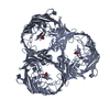
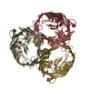
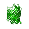
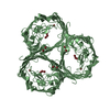
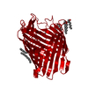
| Entry | Database: PDB / ID: 1bt9 | ||||||
|---|---|---|---|---|---|---|---|
| Title | OMPF PORIN MUTANT D74A | ||||||
 Components Components | PROTEIN (MATRIX PORIN OUTER MEMBRANE PROTEIN F) | ||||||
 Keywords Keywords | MEMBRANE PROTEIN / INTEGRAL MEMBRANE PROTEIN PORIN / PORIN | ||||||
| Function / homology |  Function and homology information Function and homology informationcolicin transmembrane transporter activity / porin activity / monoatomic ion channel complex / protein homotrimerization / pore complex / monoatomic ion channel activity / cell outer membrane / lipopolysaccharide binding / disordered domain specific binding / protein transport ...colicin transmembrane transporter activity / porin activity / monoatomic ion channel complex / protein homotrimerization / pore complex / monoatomic ion channel activity / cell outer membrane / lipopolysaccharide binding / disordered domain specific binding / protein transport / monoatomic ion transmembrane transport / lipid binding / membrane / identical protein binding Similarity search - Function | ||||||
| Biological species |  | ||||||
| Method |  X-RAY DIFFRACTION / OTHER / Resolution: 3 Å X-RAY DIFFRACTION / OTHER / Resolution: 3 Å | ||||||
 Authors Authors | Philippsen, A. / Schirmer, T. | ||||||
 Citation Citation | Journal: Biochemistry / Year: 1998 Title: Stability of trimeric OmpF porin: the contributions of the latching loop L2. Authors: Phale, P.S. / Philippsen, A. / Kiefhaber, T. / Koebnik, R. / Phale, V.P. / Schirmer, T. / Rosenbusch, J.P. #1: Journal: Nature / Year: 1992Title: Crystal Structures Explain Functional Properties of Two E.Coli Porins Authors: Cowan, S.W. / Schirmer, T. / Rummel, G. / Steiert, M. / Ghosh, R. / Pauptit, R.A. / Jansonius, J.N. / Rosenbusch, J.P. | ||||||
| History |
|
- Structure visualization
| Structure viewer | Molecule: MolmilJmol/JSmol |
|---|
- Downloads & links
-Download
| PDBx/mmCIF format | 1bt9.cif.gz | 76.8 KB | Display | PDBx/mmCIF format |
|---|---|---|---|---|
| PDB format | pdb1bt9.ent.gz | 58.3 KB | Display | PDB format |
| PDBx/mmJSON format | 1bt9.json.gz | Tree view | PDBx/mmJSON format | |
| Others |  Other downloads Other downloads |
-Validation report
| Arichive directory | https://data.pdbj.org/pub/pdb/validation_reports/bt/1bt9ftp://data.pdbj.org/pub/pdb/validation_reports/bt/1bt9 | HTTPS FTP |
|---|
-Related structure data
| Similar structure data |
|---|


-Links
 PDBj
PDBj- Assembly
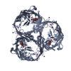
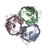
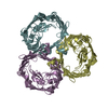
| Deposited unit | 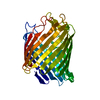
| ||||||||
|---|---|---|---|---|---|---|---|---|---|
| 1 | 
| ||||||||
| Unit cell |
|
-Components
| #1: Protein | Mass: 37070.242 Da / Num. of mol.: 1 / Mutation: D74A Source method: isolated from a genetically manipulated source Source: (gene. exp.) |
|---|---|
| #2: Water | ChemComp-HOH /  Mass: 18.015 Da / Num. of mol.: 16 / Source method: isolated from a natural source / Formula: H2O Mass: 18.015 Da / Num. of mol.: 16 / Source method: isolated from a natural source / Formula: H2O |
-Experimental details
-Experiment
| Experiment | Method: X-RAY DIFFRACTION / Number of used crystals: 1 |
|---|
- Sample preparation
| Crystal | Density Matthews: 2.88 Å3/Da / Density % sol: 57 % | |||||||||||||||
|---|---|---|---|---|---|---|---|---|---|---|---|---|---|---|---|---|
| Crystal grow | pH: 7 / Details: pH 7.0 | |||||||||||||||
| Crystal | *PLUS | |||||||||||||||
| Crystal grow | *PLUS Method: microdialysis / Details: Pauptit, R.A., (1991) J. Mol. Biol., 218, 505. | |||||||||||||||
| Components of the solutions | *PLUS
|
-Data collection
| Diffraction | Mean temperature: 276 K |
|---|---|
| Diffraction source | Wavelength: 1.5418 |
| Detector | Type: MARRESEARCH / Detector: IMAGE PLATE |
| Radiation | Protocol: SINGLE WAVELENGTH / Monochromatic (M) / Laue (L): M / Scattering type: x-ray |
| Radiation wavelength | Wavelength: 1.5418 Å / Relative weight: 1 |
| Reflection | Resolution: 3→20 Å / Num. obs: 8162 / % possible obs: 92 % / Redundancy: 3.9 % / Rmerge(I) obs: 0.082 |
- Processing
| Software |
| |||||||||||||||||||||||||||||||||||||||||||||||||||||||||||||||
|---|---|---|---|---|---|---|---|---|---|---|---|---|---|---|---|---|---|---|---|---|---|---|---|---|---|---|---|---|---|---|---|---|---|---|---|---|---|---|---|---|---|---|---|---|---|---|---|---|---|---|---|---|---|---|---|---|---|---|---|---|---|---|---|---|
| Refinement | Method to determine structure: OTHER / Resolution: 3→20 Å / Cross valid method: THROUGHOUT / σ(F): 0
| |||||||||||||||||||||||||||||||||||||||||||||||||||||||||||||||
| Refinement step | Cycle: LAST / Resolution: 3→20 Å
| |||||||||||||||||||||||||||||||||||||||||||||||||||||||||||||||
| Refine LS restraints |
| |||||||||||||||||||||||||||||||||||||||||||||||||||||||||||||||
| Software | *PLUS Name: REFMAC / Classification: refinement | |||||||||||||||||||||||||||||||||||||||||||||||||||||||||||||||
| Refinement | *PLUS Highest resolution: 3 Å / σ(F): 0 / % reflection Rfree: 10 % / Rfactor obs: 0.191 | |||||||||||||||||||||||||||||||||||||||||||||||||||||||||||||||
| Solvent computation | *PLUS | |||||||||||||||||||||||||||||||||||||||||||||||||||||||||||||||
| Displacement parameters | *PLUS |
