 Movie
Movie Controller

[English] 日本語
 Yorodumi
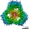
- PDB-6rd3: Crystal structure of the wild type OmpK36 from Klebsiella pneumonia -
+ Open data

- Basic information
| Entry | Database: PDB / ID: 6rd3 | ||||||
|---|---|---|---|---|---|---|---|
| Title | Crystal structure of the wild type OmpK36 from Klebsiella pneumonia | ||||||
 Components Components | OmpK36 | ||||||
 Keywords Keywords | MEMBRANE PROTEIN / OmpK36 WT | ||||||
| Function / homology |  Function and homology information Function and homology informationporin activity / pore complex / cell outer membrane / monoatomic ion transmembrane transport / metal ion binding Similarity search - Function | ||||||
| Biological species |  Klebsiella pneumoniae (bacteria) Klebsiella pneumoniae (bacteria) | ||||||
| Method |  X-RAY DIFFRACTION / SYNCHROTRON / MOLECULAR REPLACEMENT / Resolution: 1.98 Å X-RAY DIFFRACTION / SYNCHROTRON / MOLECULAR REPLACEMENT / Resolution: 1.98 Å | ||||||
 Authors Authors | Beis, K. / Romano, M. / Kwong, J. | ||||||
 Citation Citation | Journal: Nat Commun / Year: 2019 Title: OmpK36-mediated Carbapenem resistance attenuates ST258 Klebsiella pneumoniae in vivo. Authors: Wong, J.L.C. / Romano, M. / Kerry, L.E. / Kwong, H.S. / Low, W.W. / Brett, S.J. / Clements, A. / Beis, K. / Frankel, G. | ||||||
| History |
|
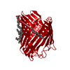
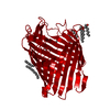
- Structure visualization
| Structure viewer | Molecule: MolmilJmol/JSmol |
|---|
- Downloads & links
-Download
| PDBx/mmCIF format | 6rd3.cif.gz | 427.3 KB | Display | PDBx/mmCIF format |
|---|---|---|---|---|
| PDB format | pdb6rd3.ent.gz | 344.3 KB | Display | PDB format |
| PDBx/mmJSON format | 6rd3.json.gz | Tree view | PDBx/mmJSON format | |
| Others |  Other downloads Other downloads |
-Validation report
| Arichive directory | https://data.pdbj.org/pub/pdb/validation_reports/rd/6rd3ftp://data.pdbj.org/pub/pdb/validation_reports/rd/6rd3 | HTTPS FTP |
|---|
-Related structure data
| Related structure data |  6rckC  6rcpC  5o79S S: Starting model for refinement C: citing same article ( |
|---|---|
| Similar structure data |


-Links
 PDBj
PDBj- Assembly
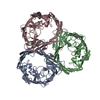
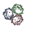
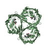
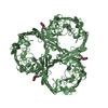
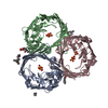
| Deposited unit | 
| ||||||||
|---|---|---|---|---|---|---|---|---|---|
| 1 | 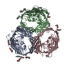
| ||||||||
| 2 | 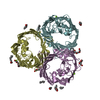
| ||||||||
| Unit cell |
|
-Components
| #1: Protein | Mass: 38058.918 Da / Num. of mol.: 6 / Mutation: G0 Source method: isolated from a genetically manipulated source Details: G0 is from cloning / Source: (gene. exp.) Klebsiella pneumoniae (bacteria) / Gene: ompK36 / Production host: #2: Chemical | ChemComp-C8E / (   Mass: 306.438 Da / Num. of mol.: 29 / Source method: obtained synthetically / Formula: C16H34O5 / Comment: C8E, detergent*YM Mass: 306.438 Da / Num. of mol.: 29 / Source method: obtained synthetically / Formula: C16H34O5 / Comment: C8E, detergent*YM#3: Chemical | ChemComp-MG /   Mass: 24.305 Da / Num. of mol.: 6 / Source method: obtained synthetically / Formula: Mg Mass: 24.305 Da / Num. of mol.: 6 / Source method: obtained synthetically / Formula: Mg#4: Water | ChemComp-HOH / |  Mass: 18.015 Da / Num. of mol.: 882 / Source method: isolated from a natural source / Formula: H2O Mass: 18.015 Da / Num. of mol.: 882 / Source method: isolated from a natural source / Formula: H2O |
|---|
-Experimental details
-Experiment
| Experiment | Method: X-RAY DIFFRACTION / Number of used crystals: 1 |
|---|
- Sample preparation
| Crystal | Density Matthews: 2.77 Å3/Da / Density % sol: 55.52 % |
|---|---|
| Crystal grow | Temperature: 293 K / Method: vapor diffusion, sitting drop Details: 0.1M Lithium sulfate, 0.1M sodium citrate pH 5.6 and 12% PEG4000 |
-Data collection
| Diffraction | Mean temperature: 100 K / Serial crystal experiment: N |
|---|---|
| Diffraction source | Source: SYNCHROTRON / Site: Diamond  / Beamline: I04 / Wavelength: 0.97 Å / Beamline: I04 / Wavelength: 0.97 Å |
| Detector | Type: DECTRIS PILATUS 6M-F / Detector: PIXEL / Date: Aug 8, 2018 |
| Radiation | Protocol: SINGLE WAVELENGTH / Monochromatic (M) / Laue (L): M / Scattering type: x-ray |
| Radiation wavelength | Wavelength: 0.97 Å / Relative weight: 1 |
| Reflection | Resolution: 1.98→53.2 Å / Num. obs: 169857 / % possible obs: 100 % / Redundancy: 6.9 % / CC1/2: 0.99 / Rmerge(I) obs: 0.023 / Net I/σ(I): 6.3 |
| Reflection shell | Resolution: 1.98→2.01 Å / Rmerge(I) obs: 0.134 / Num. unique obs: 8440 / CC1/2: 0.5 |
- Processing
| Software |
| |||||||||||||||||||||||||||||||||||||||||||||||||||||||||||||||||||||||||||||||||||||||||||||||||||||||||||||||||||||||||||||||||||||||||||||||||||||||||||||||||||||||||||||||||||||||||||||||||||||||||||||||||||||||||
|---|---|---|---|---|---|---|---|---|---|---|---|---|---|---|---|---|---|---|---|---|---|---|---|---|---|---|---|---|---|---|---|---|---|---|---|---|---|---|---|---|---|---|---|---|---|---|---|---|---|---|---|---|---|---|---|---|---|---|---|---|---|---|---|---|---|---|---|---|---|---|---|---|---|---|---|---|---|---|---|---|---|---|---|---|---|---|---|---|---|---|---|---|---|---|---|---|---|---|---|---|---|---|---|---|---|---|---|---|---|---|---|---|---|---|---|---|---|---|---|---|---|---|---|---|---|---|---|---|---|---|---|---|---|---|---|---|---|---|---|---|---|---|---|---|---|---|---|---|---|---|---|---|---|---|---|---|---|---|---|---|---|---|---|---|---|---|---|---|---|---|---|---|---|---|---|---|---|---|---|---|---|---|---|---|---|---|---|---|---|---|---|---|---|---|---|---|---|---|---|---|---|---|---|---|---|---|---|---|---|---|---|---|---|---|---|---|---|---|
| Refinement | Method to determine structure: MOLECULAR REPLACEMENT Starting model: 5O79 Resolution: 1.98→53.197 Å / SU ML: 0.22 / Cross valid method: FREE R-VALUE / σ(F): 1.35 / Phase error: 23.27 / Stereochemistry target values: ML
| |||||||||||||||||||||||||||||||||||||||||||||||||||||||||||||||||||||||||||||||||||||||||||||||||||||||||||||||||||||||||||||||||||||||||||||||||||||||||||||||||||||||||||||||||||||||||||||||||||||||||||||||||||||||||
| Solvent computation | Shrinkage radii: 0.9 Å / VDW probe radii: 1.11 Å / Solvent model: FLAT BULK SOLVENT MODEL | |||||||||||||||||||||||||||||||||||||||||||||||||||||||||||||||||||||||||||||||||||||||||||||||||||||||||||||||||||||||||||||||||||||||||||||||||||||||||||||||||||||||||||||||||||||||||||||||||||||||||||||||||||||||||
| Refinement step | Cycle: LAST / Resolution: 1.98→53.197 Å
| |||||||||||||||||||||||||||||||||||||||||||||||||||||||||||||||||||||||||||||||||||||||||||||||||||||||||||||||||||||||||||||||||||||||||||||||||||||||||||||||||||||||||||||||||||||||||||||||||||||||||||||||||||||||||
| Refine LS restraints |
| |||||||||||||||||||||||||||||||||||||||||||||||||||||||||||||||||||||||||||||||||||||||||||||||||||||||||||||||||||||||||||||||||||||||||||||||||||||||||||||||||||||||||||||||||||||||||||||||||||||||||||||||||||||||||
| LS refinement shell |
|
