+ Open data
- Basic information
| Entry | Database: EMDB / ID: EMD-10754 | |||||||||
|---|---|---|---|---|---|---|---|---|---|---|
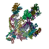
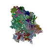
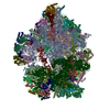
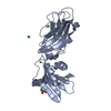
| Title | Clathrin with bound beta2 appendage of AP2 | |||||||||
 Map data Map data | Sharpened map of hexagon bounding clathrin leg enriched in beta-appendage | |||||||||
 Sample Sample |
| |||||||||
 Keywords Keywords | clathrin / clathrin adaptor / ap2 / clathrin assembly / ENDOCYTOSIS | |||||||||
| Function / homology |  Function and homology information Function and homology informationLysosome Vesicle Biogenesis / WNT5A-dependent internalization of FZD4 / WNT5A-dependent internalization of FZD2, FZD5 and ROR2 / Cargo recognition for clathrin-mediated endocytosis / Gap junction degradation / Formation of annular gap junctions / Clathrin-mediated endocytosis / clathrin coat of trans-Golgi network vesicle / clathrin light chain binding / clathrin complex ...Lysosome Vesicle Biogenesis / WNT5A-dependent internalization of FZD4 / WNT5A-dependent internalization of FZD2, FZD5 and ROR2 / Cargo recognition for clathrin-mediated endocytosis / Gap junction degradation / Formation of annular gap junctions / Clathrin-mediated endocytosis / clathrin coat of trans-Golgi network vesicle / clathrin light chain binding / clathrin complex / Nef Mediated CD8 Down-regulation / clathrin adaptor complex / WNT5A-dependent internalization of FZD2, FZD5 and ROR2 / Trafficking of GluR2-containing AMPA receptors / WNT5A-dependent internalization of FZD4 / clathrin coat of coated pit / AP-2 adaptor complex / postsynaptic neurotransmitter receptor internalization / LDL clearance / clathrin coat assembly / Retrograde neurotrophin signalling / clathrin-coated endocytic vesicle / clathrin-dependent endocytosis / coronary vasculature development / endolysosome membrane / signal sequence binding / Nef Mediated CD4 Down-regulation / aorta development / clathrin-coated vesicle / ventricular septum development / ciliary membrane / clathrin binding / Recycling pathway of L1 / synaptic vesicle endocytosis / EPH-ephrin mediated repulsion of cells / vesicle-mediated transport / MHC class II antigen presentation / VLDLR internalisation and degradation / receptor-mediated endocytosis / kidney development / intracellular protein transport / clathrin-coated endocytic vesicle membrane / trans-Golgi network / cytoplasmic side of plasma membrane / spindle / disordered domain specific binding / endocytic vesicle membrane / Cargo recognition for clathrin-mediated endocytosis / Clathrin-mediated endocytosis / presynapse / mitotic cell cycle / postsynapse / Potential therapeutics for SARS / glutamatergic synapse / nucleolus / structural molecule activity / extracellular exosome / nucleoplasm / membrane / plasma membrane / cytosol Similarity search - Function | |||||||||
| Biological species |   Homo sapiens (human) Homo sapiens (human) | |||||||||
| Method | subtomogram averaging / cryo EM / Resolution: 9.2 Å | |||||||||
 Authors Authors | Kovtun O / Kane Dickson V | |||||||||
| Funding support |  United Kingdom, 2 items United Kingdom, 2 items
| |||||||||
 Citation Citation |  Journal: Sci Adv / Year: 2020 Journal: Sci Adv / Year: 2020Title: Architecture of the AP2/clathrin coat on the membranes of clathrin-coated vesicles. Authors: Oleksiy Kovtun / Veronica Kane Dickson / Bernard T Kelly / David J Owen / John A G Briggs /   Abstract: Clathrin-mediated endocytosis (CME) is crucial for modulating the protein composition of a cell's plasma membrane. Clathrin forms a cage-like, polyhedral outer scaffold around a vesicle, to which ...Clathrin-mediated endocytosis (CME) is crucial for modulating the protein composition of a cell's plasma membrane. Clathrin forms a cage-like, polyhedral outer scaffold around a vesicle, to which cargo-selecting clathrin adaptors are attached. Adaptor protein complex (AP2) is the key adaptor in CME. Crystallography has shown AP2 to adopt a range of conformations. Here, we used cryo-electron microscopy, tomography, and subtomogram averaging to determine structures, interactions, and arrangements of clathrin and AP2 at the key steps of coat assembly, from AP2 in solution to membrane-assembled clathrin-coated vesicles (CCVs). AP2 binds cargo and PtdIns(4,5) (phosphatidylinositol 4,5-bisphosphate)-containing membranes via multiple interfaces, undergoing conformational rearrangement from its cytosolic state. The binding mode of AP2 β2 appendage into the clathrin lattice in CCVs and buds implies how the adaptor structurally modulates coat curvature and coat disassembly. | |||||||||
| History |
|
- Structure visualization
| Movie |
 Movie viewer Movie viewer |
|---|---|
| Structure viewer | EM map:  SurfView SurfView Molmil Molmil Jmol/JSmol Jmol/JSmol |
| Supplemental images |
- Downloads & links
-EMDB archive
| Map data |  emd_10754.map.gz emd_10754.map.gz | 9.8 MB |  EMDB map data format EMDB map data format | |
|---|---|---|---|---|
| Header (meta data) |  emd-10754-v30.xml emd-10754-v30.xml emd-10754.xml emd-10754.xml | 30 KB 30 KB | Display Display |  EMDB header EMDB header |
| FSC (resolution estimation) |  emd_10754_fsc.xml emd_10754_fsc.xml | 5.8 KB | Display |  FSC data file FSC data file |
| Images |  emd_10754.png emd_10754.png | 315.3 KB | ||
| Masks |  emd_10754_msk_1.map emd_10754_msk_1.map | 15.6 MB |  Mask map Mask map | |
| Filedesc metadata |  emd-10754.cif.gz emd-10754.cif.gz | 8.6 KB | ||
| Others |  emd_10754_half_map_1.map.gz emd_10754_half_map_1.map.gz emd_10754_half_map_2.map.gz emd_10754_half_map_2.map.gz | 14.4 MB 14.5 MB | ||
| Archive directory |  http://ftp.pdbj.org/pub/emdb/structures/EMD-10754 http://ftp.pdbj.org/pub/emdb/structures/EMD-10754 ftp://ftp.pdbj.org/pub/emdb/structures/EMD-10754 ftp://ftp.pdbj.org/pub/emdb/structures/EMD-10754 | HTTPS FTP |
-Validation report
| Summary document |  emd_10754_validation.pdf.gz emd_10754_validation.pdf.gz | 942.7 KB | Display |  EMDB validaton report EMDB validaton report |
|---|---|---|---|---|
| Full document |  emd_10754_full_validation.pdf.gz emd_10754_full_validation.pdf.gz | 494.5 KB | Display | |
| Data in XML |  emd_10754_validation.xml.gz emd_10754_validation.xml.gz | 11.6 KB | Display | |
| Data in CIF |  emd_10754_validation.cif.gz emd_10754_validation.cif.gz | 15.6 KB | Display | |
| Arichive directory |  https://ftp.pdbj.org/pub/emdb/validation_reports/EMD-10754 https://ftp.pdbj.org/pub/emdb/validation_reports/EMD-10754 ftp://ftp.pdbj.org/pub/emdb/validation_reports/EMD-10754 ftp://ftp.pdbj.org/pub/emdb/validation_reports/EMD-10754 | HTTPS FTP |
-Related structure data
| Related structure data |  6yaiMC  6yaeC  6yafC  6yahC C: citing same article ( M: atomic model generated by this map |
|---|---|
| Similar structure data |
- Links
Links
| EMDB pages |  EMDB (EBI/PDBe) / EMDB (EBI/PDBe) /  EMDataResource EMDataResource |
|---|---|
| Related items in Molecule of the Month |
- Map
Map
| File |  Download / File: emd_10754.map.gz / Format: CCP4 / Size: 15.6 MB / Type: IMAGE STORED AS FLOATING POINT NUMBER (4 BYTES) Download / File: emd_10754.map.gz / Format: CCP4 / Size: 15.6 MB / Type: IMAGE STORED AS FLOATING POINT NUMBER (4 BYTES) | ||||||||||||||||||||||||||||||||||||||||||||||||||||||||||||
|---|---|---|---|---|---|---|---|---|---|---|---|---|---|---|---|---|---|---|---|---|---|---|---|---|---|---|---|---|---|---|---|---|---|---|---|---|---|---|---|---|---|---|---|---|---|---|---|---|---|---|---|---|---|---|---|---|---|---|---|---|---|
| Annotation | Sharpened map of hexagon bounding clathrin leg enriched in beta-appendage | ||||||||||||||||||||||||||||||||||||||||||||||||||||||||||||
| Projections & slices | Image control
Images are generated by Spider. | ||||||||||||||||||||||||||||||||||||||||||||||||||||||||||||
| Voxel size | X=Y=Z: 1.78 Å | ||||||||||||||||||||||||||||||||||||||||||||||||||||||||||||
| Density |
| ||||||||||||||||||||||||||||||||||||||||||||||||||||||||||||
| Symmetry | Space group: 1 | ||||||||||||||||||||||||||||||||||||||||||||||||||||||||||||
| Details | EMDB XML:
CCP4 map header:
| ||||||||||||||||||||||||||||||||||||||||||||||||||||||||||||
-Supplemental data
-Mask #1
| File |  emd_10754_msk_1.map emd_10754_msk_1.map | ||||||||||||
|---|---|---|---|---|---|---|---|---|---|---|---|---|---|
| Projections & Slices |
| ||||||||||||
| Density Histograms |
-Half map: Unsharpened half-map of hexagon bounding clathrin leg enriched...
| File | emd_10754_half_map_1.map | ||||||||||||
|---|---|---|---|---|---|---|---|---|---|---|---|---|---|
| Annotation | Unsharpened half-map of hexagon bounding clathrin leg enriched in beta2-appendage | ||||||||||||
| Projections & Slices |
| ||||||||||||
| Density Histograms |
-Half map: Unsharpened half-map of hexagon bounding clathrin leg enriched...
| File | emd_10754_half_map_2.map | ||||||||||||
|---|---|---|---|---|---|---|---|---|---|---|---|---|---|
| Annotation | Unsharpened half-map of hexagon bounding clathrin leg enriched in beta-appendage | ||||||||||||
| Projections & Slices |
| ||||||||||||
| Density Histograms |
- Sample components
-Entire : Clathrin/AP2 coat assembled on membrane.
| Entire | Name: Clathrin/AP2 coat assembled on membrane. |
|---|---|
| Components |
|
-Supramolecule #1: Clathrin/AP2 coat assembled on membrane.
| Supramolecule | Name: Clathrin/AP2 coat assembled on membrane. / type: complex / ID: 1 / Parent: 0 / Macromolecule list: all Details: Clathrin/AP coat was formed on the membrane containing cargo signal peptides. The AP2 adaptor lacked hinge and appendage regions in its alpha subunit. |
|---|
-Supramolecule #2: Clathrin
| Supramolecule | Name: Clathrin / type: complex / ID: 2 / Parent: 1 / Macromolecule list: #1, #3-#4 Details: Clathrin in the clathrin/AP2 coat formed on the membrane |
|---|---|
| Source (natural) | Organism:  |
-Supramolecule #3: beta2 appendage of AP2 adaptor
| Supramolecule | Name: beta2 appendage of AP2 adaptor / type: complex / ID: 3 / Parent: 1 / Macromolecule list: #2 Details: beta2 appendage of the AP2 bound to clathrin to clathrin/AP2 coat formed on the membrane |
|---|---|
| Source (natural) | Organism:  Homo sapiens (human) Homo sapiens (human) |
-Macromolecule #1: Clathrin heavy chain
| Macromolecule | Name: Clathrin heavy chain / type: protein_or_peptide / ID: 1 / Number of copies: 8 / Enantiomer: LEVO |
|---|---|
| Source (natural) | Organism:  |
| Molecular weight | Theoretical: 187.145125 KDa |
| Sequence | String: MAQILPIRFQ EHLQLQNLGI NPANIGFSTL TMESDKFICI REKVGEQAQV VIIDMNDPSN PIRRPISADS AIMNPASKVI ALKAGKTLQ IFNIEMKSKM KAHTMTDDVT FWKWISLNTV ALVTDNAVYH WSMEGESQPV KMFDRHSSLA GCQIINYRTD A KQKWLLLT ...String: MAQILPIRFQ EHLQLQNLGI NPANIGFSTL TMESDKFICI REKVGEQAQV VIIDMNDPSN PIRRPISADS AIMNPASKVI ALKAGKTLQ IFNIEMKSKM KAHTMTDDVT FWKWISLNTV ALVTDNAVYH WSMEGESQPV KMFDRHSSLA GCQIINYRTD A KQKWLLLT GISAQQNRVV GAMQLYSVDR KVSQPIEGHA ASFAQFKMEG NAEESTLFCF AVRGQAGGKL HIIEVGTPPT GN QPFPKKA VDVFFPPEAQ NDFPVAMQIS EKHDVVFLIT KYGYIHLYDL ETGTCIYMNR ISGETIFVTA PHEATAGIIG VNR KGQVLS VCVEEENIIP YITNVLQNPD LALRMAVRNN LAGAEELFAR KFNALFAQGN YSEAAKVAAN APKGILRTPD TIRR FQSVP AQPGQTSPLL QYFGILLDQG QLNKYESLEL CRPVLQQGRK QLLEKWLKED KLECSEELGD LVKSVDPTLA LSVYL RANV PNKVIQCFAE TGQVQKIVLY AKKVGYTPDW IFLLRNVMRI SPDQGQQFAQ MLVQDEEPLA DITQIVDVFM EYNLIQ QCT AFLLDALKNN RPSEGPLQTR LLEMNLMHAP QVADAILGNQ MFTHYDRAHI AQLCEKAGLL QRALEHFTDL YDIKRAV VH THLLNPEWLV NYFGSLSVED SLECLRAMLS ANIRQNLQIC VQVASKYHEQ LSTQSLIELF ESFKSFEGLF YFLGSIVN F SQDPDVHFKY IQAACKTGQI KEVERICRES NCYDPERVKN FLKEAKLTDQ LPLIIVCDRF DFVHDLVLYL YRNNLQKYI EIYVQKVNPS RLPVVIGGLL DVDCSEDVIK NLILVVRGQF STDELVAEVE KRNRLKLLLP WLEARIHEGC EEPATHNALA KIYIDSNNN PERFLRENPY YDSRVVGKYC EKRDPHLACV AYERGQCDLE LINVCNENSL FKSLSRYLVR RKDPELWGSV L LESNPYRR PLIDQVVQTA LSETQDPEEV SVTVKAFMTA DLPNELIELL EKIVLDNSVF SEHRNLQNLL ILTAIKADRT RV MEYINRL DNYDAPDIAN IAISNELFEE AFAIFRKFDV NTSAVQVLIE HIGNLDRAYE FAERCNEPAV WSQLAKAQLQ KGM VKEAID SYIKADDPSS YMEVVQAANT SGNWEELVKY LQMARKKARE SYVETELIFA LAKTNRLAEL EEFINGPNNA HIQQ VGDRC YDEKMYDAAK LLYNNVSNFG RLASTLVHLG EYQAAVDGAR KANSTRTWKE VCFACVDGKE FRLAQMCGLH IVVHA DELE ELINYYQDRG YFEELITMLE AALGLERAHM GMFTELAILY SKFKPQKMRE HLELFWSRVN IPKVLRAAEQ AHLWAE LVF LYDKYEEYDN AIITMMNHPT DAWKEGQFKD IITKVANVEL YYRAIQFYLE FKPLLLNDLL MVLSPRLDHT RAVNYFS KV KQLPLVKPYL RSVQNHNNKS VNESLNNLFI TEEDYQALRT SIDAYDNFDN ISLAQRLEKH ELIEFRRIAA YLFKGNNR W KQSVELCKKD SLYKDAMQYA SESKDTELAE ELLQWFLQEE KRECFGACLF TCYDLLRPDV VLETAWRHNI MDFAMPYFI QVMKEYLTKV DKLDASESLR KEEEQATETQ UniProtKB: Clathrin heavy chain |
-Macromolecule #2: AP-2 complex subunit beta
| Macromolecule | Name: AP-2 complex subunit beta / type: protein_or_peptide / ID: 2 / Number of copies: 1 / Enantiomer: LEVO |
|---|---|
| Source (natural) | Organism:  Homo sapiens (human) Homo sapiens (human) |
| Molecular weight | Theoretical: 26.429457 KDa |
| Recombinant expression | Organism:  |
| Sequence | String: GGYVAPKAVW LPAVKAKGLE ISGTFTHRQG HIYMEMNFTN KALQHMTDFA IQFNKNSFGV IPSTPLAIHT PLMPNQSIDV SLPLNTLGP VMKMEPLNNL QVAVKNNIDV FYFSCLIPLN VLFVEDGKME RQVFLATWKD IPNENELQFQ IKECHLNADT V SSKLQNNN ...String: GGYVAPKAVW LPAVKAKGLE ISGTFTHRQG HIYMEMNFTN KALQHMTDFA IQFNKNSFGV IPSTPLAIHT PLMPNQSIDV SLPLNTLGP VMKMEPLNNL QVAVKNNIDV FYFSCLIPLN VLFVEDGKME RQVFLATWKD IPNENELQFQ IKECHLNADT V SSKLQNNN VYTIAKRNVE GQDMLYQSLK LTNGIWILAE LRIQPGNPNY TLSLKCRAPE VSQYIYQVYD SILKN UniProtKB: AP-2 complex subunit beta |
-Macromolecule #3: Clathrin heavy chain
| Macromolecule | Name: Clathrin heavy chain / type: protein_or_peptide / ID: 3 / Number of copies: 1 / Enantiomer: LEVO |
|---|---|
| Source (natural) | Organism:  |
| Molecular weight | Theoretical: 187.117094 KDa |
| Sequence | String: MAQILPIRFQ EHLQLQNLGI NPANIGFSTL TMESDKFICI REKVGEQAQV VIIDMNDPSN PIRRPISADS AIMNPASKVI ALKAGKTLQ IFNIEMKSKM KAHTMTDDVT FWKWISLNTV ALVTDNAVYH WSMEGESQPV KMFDRHSSLA GCQIINYRTD A KQKWLLLT ...String: MAQILPIRFQ EHLQLQNLGI NPANIGFSTL TMESDKFICI REKVGEQAQV VIIDMNDPSN PIRRPISADS AIMNPASKVI ALKAGKTLQ IFNIEMKSKM KAHTMTDDVT FWKWISLNTV ALVTDNAVYH WSMEGESQPV KMFDRHSSLA GCQIINYRTD A KQKWLLLT GISAQQNRVV GAMQLYSVDR KVSQPIEGHA ASFAQFKMEG NAEESTLFCF AVRGQAGGKL HIIEVGTPPT GN QPFPKKA VDVFFPPEAQ NDFPVAMQIS EKHDVVFLIT KYGYIHLYDL ETGTCIYMNR ISGETIFVTA PHEATAGIIG VNR KGQVLS VCVEEENIIP YITNVLQNPD LALRMAVRNN LAGAEELFAR KFNALFAQGN YSEAAKVAAN APKGILRTPD TIRR FQSVP AQPGQTSPLL QYFGILLDQG QLNKYESLEL CRPVLQQGRK QLLEKWLKED KLECSEELGD LVKSVDPTLA LSVYL RANV PNKVIQCFAE TGQVQKIVLY AKKVGYTPDW IFLLRNVMRI SPDQGQQFAQ MLVQDEEPLA DITQIVDVFM EYNLIQ QCT AFLLDALKNN RPSEGPLQTR LLEMNLMHAP QVADAILGNQ MFTHYDRAHI AQLCEKAGLL QRALEHFTDL YDIKRAV VH THLLNPEWLV NYFGSLSVED SLECLRAMLS ANIRQNLQIC VQVASKYHEQ LSTQSLIELF ESFKSFEGLF YFLGSIVN F SQDPDVHFKY IQAACKTGQI KEVERICRES NCYDPERVKN FLKEAKLTDQ LPLIIVCDRF DFVHDLVLYL YRNNLQKYI EIYVQKVNPS RLPVVIGGLL DVDCSEDVIK NLILVVRGQF STDELVAEVE KRNRLKLLLP WLEARIHEGC TEPATHNALA KIYIDSNNN PERFLRENPY YDSRVVGKYC EKRDPHLACV AYERGQCDLE LINVCNENSL FKSLSRYLVR RKDPELWGSV L LESNPYRR PLIDQVVQTA LSETQDPEEV SVTVKAFMTA DLPNELIELL EKIVLDNSVF SEHRNLQNLL ILTAIKADRT RV MEYINRL DNYDAPDIAN IAISNELFEE AFAIFRKFDV NTSAVQVLIE HIGNLDRAYE FAERCNEPAV WSQLAKAQLQ KGM VKEAID SYIKADDPSS YMEVVQAANT SGNWEELVKY LQMARKKARE SYVETELIFA LAKTNRLAEL EEFINGPNNA HIQQ VGDRC YDEKMYDAAK LLYNNVSNFG RLASTLVHLG EYQAAVDGAR KANSTRTWKE VCFACVDGKE FRLAQMCGLH IVVHA DELE ELINYYQDRG YFEELITMLE AALGLERAHM GMFTELAILY SKFKPQKMRE HLELFWSRVN IPKVLRAAEQ AHLWAE LVF LYDKYEEYDN AIITMMNHPT DAWKEGQFKD IITKVANVEL YYRAIQFYLE FKPLLLNDLL MVLSPRLDHT RAVNYFS KV KQLPLVKPYL RSVQNHNNKS VNESLNNLFI TEEDYQALRT SIDAYDNFDN ISLAQRLEKH ELIEFRRIAA YLFKGNNR W KQSVELCKKD SLYKDAMQYA SESKDTELAE ELLQWFLQEE KRECFGACLF TCYDLLRPDV VLETAWRHNI MDFAMPYFI QVMKEYLTKV DKLDASESLR KEEEQATETQ UniProtKB: Clathrin heavy chain |
-Macromolecule #4: Clathrin light chain
| Macromolecule | Name: Clathrin light chain / type: protein_or_peptide / ID: 4 / Number of copies: 4 / Enantiomer: LEVO |
|---|---|
| Source (natural) | Organism:  |
| Molecular weight | Theoretical: 25.2185 KDa |
| Sequence | String: MADDFGFFSS SESGAPEVAE EDPAAAFLAQ QESEIAGIEN DEGFGAPAGS QAALAQPGPA SGAGPEDMGT TVNGDVFQDA NGPADGYAA IAQADRLTQE PESIRKWREE QRKRLQELDA ASKVTEQEWR EKAKKDLEEW NQRQSEQVEK NKINNRIADK A FYQQPDAD ...String: MADDFGFFSS SESGAPEVAE EDPAAAFLAQ QESEIAGIEN DEGFGAPAGS QAALAQPGPA SGAGPEDMGT TVNGDVFQDA NGPADGYAA IAQADRLTQE PESIRKWREE QRKRLQELDA ASKVTEQEWR EKAKKDLEEW NQRQSEQVEK NKINNRIADK A FYQQPDAD IIGYVASEEA FVKESKEETP GTEWEKVAQL CDFNPKSSKQ CKDVSRLRSV LMSLKQTPLS R UniProtKB: Nucleolar protein 16 |
-Experimental details
-Structure determination
| Method | cryo EM |
|---|---|
 Processing Processing | subtomogram averaging |
| Aggregation state | 3D array |
- Sample preparation
| Concentration | 0.7 mg/mL | ||||||||||
|---|---|---|---|---|---|---|---|---|---|---|---|
| Buffer | pH: 7.2 Component:
| ||||||||||
| Grid | Model: C-flat-2/2 / Support film - Material: CARBON / Support film - topology: HOLEY | ||||||||||
| Vitrification | Cryogen name: ETHANE / Chamber humidity: 98 % / Chamber temperature: 291 K / Instrument: LEICA EM GP Details: The sample was supplemented with 10 nm nanogold fiducials, and 3 ul of the mixture was backside blotted for 3 seconds.. | ||||||||||
| Details | The sample (in vitro budding reaction) contained AP2, clathrin and 400 nm extruded liposomes |
- Electron microscopy
| Microscope | FEI TITAN KRIOS |
|---|---|
| Specialist optics | Energy filter - Name: GIF Quantum LS / Energy filter - Slit width: 20 eV |
| Image recording | Film or detector model: GATAN K2 QUANTUM (4k x 4k) / Detector mode: COUNTING / Digitization - Dimensions - Width: 3838 pixel / Digitization - Dimensions - Height: 3710 pixel / Digitization - Frames/image: 1-10 / Number grids imaged: 2 / Average exposure time: 0.2 sec. / Average electron dose: 3.2 e/Å2 Details: The images were collected in movie mode at 10 frames per second |
| Electron beam | Acceleration voltage: 300 kV / Electron source:  FIELD EMISSION GUN FIELD EMISSION GUN |
| Electron optics | C2 aperture diameter: 100.0 µm / Calibrated defocus max: 6.5 µm / Calibrated defocus min: 1.5 µm / Calibrated magnification: 81000 / Illumination mode: FLOOD BEAM / Imaging mode: BRIGHT FIELD / Cs: 2.7 mm / Nominal defocus max: 6.5 µm / Nominal defocus min: 1.5 µm / Nominal magnification: 81000 |
| Sample stage | Specimen holder model: FEI TITAN KRIOS AUTOGRID HOLDER / Cooling holder cryogen: NITROGEN |
| Experimental equipment |  Model: Titan Krios / Image courtesy: FEI Company |
+ Image processing
-Atomic model buiding 1
| Initial model |
| ||||||||
|---|---|---|---|---|---|---|---|---|---|
| Refinement | Space: REAL / Protocol: FLEXIBLE FIT / Target criteria: correlation coefficient | ||||||||
| Output model |  PDB-6yai: |
 Movie
Movie Controller


 Z (Sec.)
 Y (Row.)
 X (Col.)


