 Movie
Movie Controller


+ Open data

- Basic information
| Entry | Database: EMDB / ID: EMD-10617 | |||||||||
|---|---|---|---|---|---|---|---|---|---|---|
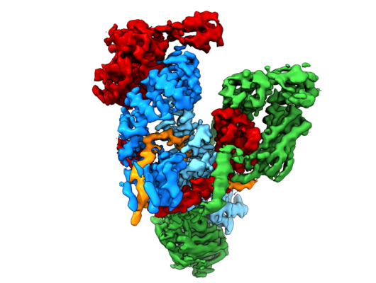
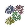
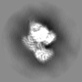
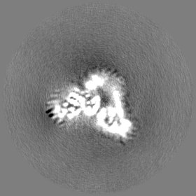
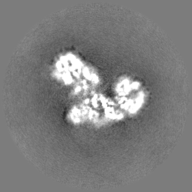
| Title | Subunits BBS 1,4,8,9,18 of the human BBSome complex | |||||||||
 Map data Map data | ||||||||||
 Sample Sample |
| |||||||||
 Keywords Keywords | ciliary transport / Arl6 effector / adaptor protein / complex / PROTEIN TRANSPORT | |||||||||
| Function / homology |  Function and homology information Function and homology informationregulation of non-motile cilium assembly / protein localization to photoreceptor outer segment / RNA polymerase II-specific DNA-binding transcription factor binding => GO:0061629 / receptor localization to non-motile cilium / BBSome / retinal rod cell development / negative regulation of appetite by leptin-mediated signaling pathway / photoreceptor cell outer segment organization / microtubule anchoring at centrosome / smoothened binding ...regulation of non-motile cilium assembly / protein localization to photoreceptor outer segment / RNA polymerase II-specific DNA-binding transcription factor binding => GO:0061629 / receptor localization to non-motile cilium / BBSome / retinal rod cell development / negative regulation of appetite by leptin-mediated signaling pathway / photoreceptor cell outer segment organization / microtubule anchoring at centrosome / smoothened binding / regulation of cilium beat frequency involved in ciliary motility / sensory processing / protein localization to organelle / ciliary transition zone / melanosome transport / photoreceptor connecting cilium / patched binding / ventricular system development / negative regulation of actin filament polymerization / BBSome-mediated cargo-targeting to cilium / striatum development / Golgi to plasma membrane protein transport / positive regulation of cilium assembly / positive regulation of multicellular organism growth / response to stimulus / non-motile cilium assembly / maintenance of protein location in nucleus / photoreceptor cell maintenance / protein localization to cilium / regulation of stress fiber assembly / brain morphogenesis / non-motile cilium / negative regulation of systemic arterial blood pressure / retina homeostasis / motile cilium / centrosome cycle / protein localization to centrosome / negative regulation of GTPase activity / ciliary membrane / fat pad development / adult behavior / pericentriolar material / beta-tubulin binding / heart looping / fat cell differentiation / dendrite development / face development / axoneme / dynactin binding / social behavior / regulation of lipid metabolic process / centriolar satellite / spermatid development / mitotic cytokinesis / photoreceptor outer segment / alpha-tubulin binding / cilium assembly / intracellular transport / visual perception / centriole / photoreceptor inner segment / ciliary basal body / regulation of cytokinesis / neural tube closure / hippocampus development / neuron migration / cerebral cortex development / cilium / microtubule cytoskeleton organization / sensory perception of smell / protein transport / protein-macromolecule adaptor activity / negative regulation of gene expression / centrosome / membrane / nucleus / cytoplasm / cytosol Similarity search - Function | |||||||||
| Biological species |  Homo sapiens (human) Homo sapiens (human) | |||||||||
| Method | single particle reconstruction / cryo EM / Resolution: 3.8 Å | |||||||||
 Authors Authors | Klink BU / Raunser S | |||||||||
| Funding support |  Germany, 1 items Germany, 1 items
| |||||||||
 Citation Citation | Journal: Elife / Year: 2020 Title: Structure of the human BBSome core complex. Authors: Björn Udo Klink / Christos Gatsogiannis / Oliver Hofnagel / Alfred Wittinghofer / Stefan Raunser / Abstract: The BBSome is a heterooctameric protein complex that plays a central role in primary cilia homeostasis. Its malfunction causes the severe ciliopathy Bardet-Biedl syndrome (BBS). The complex acts as a ...The BBSome is a heterooctameric protein complex that plays a central role in primary cilia homeostasis. Its malfunction causes the severe ciliopathy Bardet-Biedl syndrome (BBS). The complex acts as a cargo adapter that recognizes signaling proteins such as GPCRs and links them to the intraflagellar transport machinery. The underlying mechanism is poorly understood. Here we present a high-resolution cryo-EM structure of a human heterohexameric core subcomplex of the BBSome. The structure reveals the architecture of the complex in atomic detail. It explains how the subunits interact with each other and how disease-causing mutations hamper this interaction. The complex adopts a conformation that is open for binding to membrane-associated GTPase Arl6 and a large positively charged patch likely strengthens the interaction with the membrane. A prominent negatively charged cleft at the center of the complex is likely involved in binding of positively charged signaling sequences of cargo proteins. | |||||||||
| History |
|
- Structure visualization
| Movie |
Movie viewer |
|---|---|
| Structure viewer | EM map: SurfViewMolmilJmol/JSmol |
| Supplemental images |
- Downloads & links
-EMDB archive
| Map data | emd_10617.map.gz | 2.7 MB | EMDB map data format | |
|---|---|---|---|---|
| Header (meta data) | emd-10617-v30.xmlemd-10617.xml | 23.9 KB 23.9 KB | Display Display | EMDB header |
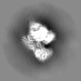
| Images |  emd_10617.png emd_10617.png | 166.1 KB | ||
| Masks | emd_10617_msk_1.map | 83.7 MB | Mask map | |
| Filedesc metadata | emd-10617.cif.gz | 8 KB | ||
| Others | emd_10617_half_map_1.map.gzemd_10617_half_map_2.map.gz | 39.4 MB 39.8 MB | ||
| Archive directory |  http://ftp.pdbj.org/pub/emdb/structures/EMD-10617ftp://ftp.pdbj.org/pub/emdb/structures/EMD-10617 http://ftp.pdbj.org/pub/emdb/structures/EMD-10617ftp://ftp.pdbj.org/pub/emdb/structures/EMD-10617 | HTTPS FTP |
-Validation report
| Summary document | emd_10617_validation.pdf.gz | 612.1 KB | Display | EMDB validaton report |
|---|---|---|---|---|
| Full document | emd_10617_full_validation.pdf.gz | 611.7 KB | Display | |
| Data in XML | emd_10617_validation.xml.gz | 12.4 KB | Display | |
| Data in CIF | emd_10617_validation.cif.gz | 14.4 KB | Display | |
| Arichive directory | https://ftp.pdbj.org/pub/emdb/validation_reports/EMD-10617ftp://ftp.pdbj.org/pub/emdb/validation_reports/EMD-10617 | HTTPS FTP |
-Related structure data
| Related structure data |  6xt9MC  6xtbC C: citing same article ( M: atomic model generated by this map |
|---|---|
| Similar structure data |


-Links
| EMDB pages | EMDB (EBI/PDBe) / EMDataResource |
|---|---|
| Related items in Molecule of the Month |


-Map
| File | Download / File: emd_10617.map.gz / Format: CCP4 / Size: 83.7 MB / Type: IMAGE STORED AS FLOATING POINT NUMBER (4 BYTES) | ||||||||||||||||||||||||||||||||||||||||||||||||||||||||||||
|---|---|---|---|---|---|---|---|---|---|---|---|---|---|---|---|---|---|---|---|---|---|---|---|---|---|---|---|---|---|---|---|---|---|---|---|---|---|---|---|---|---|---|---|---|---|---|---|---|---|---|---|---|---|---|---|---|---|---|---|---|---|
| Voxel size | X=Y=Z: 1.07 Å | ||||||||||||||||||||||||||||||||||||||||||||||||||||||||||||
| Density |
| ||||||||||||||||||||||||||||||||||||||||||||||||||||||||||||
| Symmetry | Space group: 1 | ||||||||||||||||||||||||||||||||||||||||||||||||||||||||||||
| Details | EMDB XML:
CCP4 map header:
| ||||||||||||||||||||||||||||||||||||||||||||||||||||||||||||
-Supplemental data
-Mask #1
| File | emd_10617_msk_1.map | ||||||||||||
|---|---|---|---|---|---|---|---|---|---|---|---|---|---|
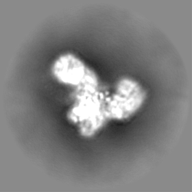
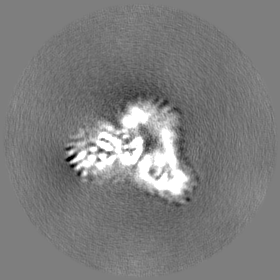
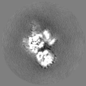
| Projections & Slices |
| ||||||||||||
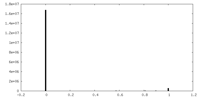
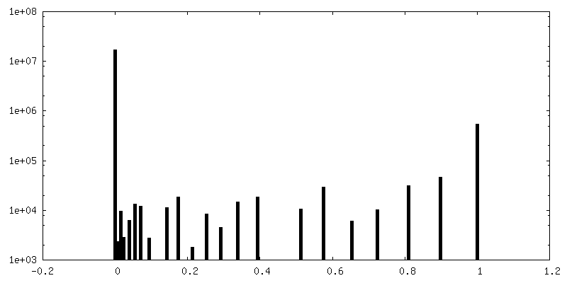
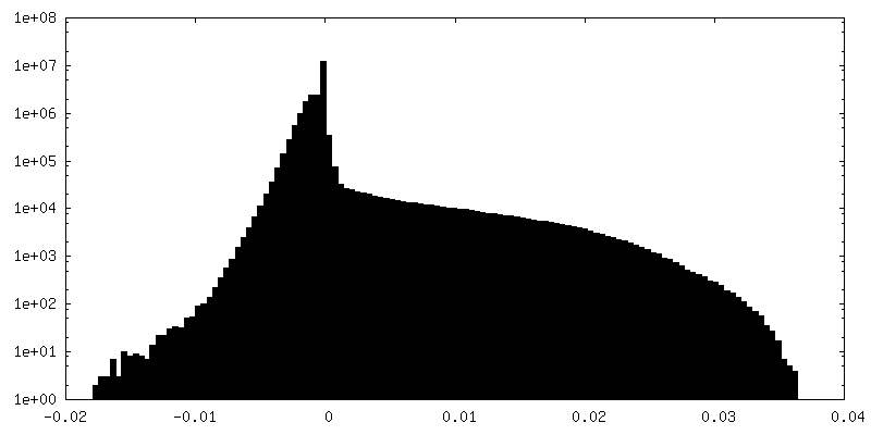
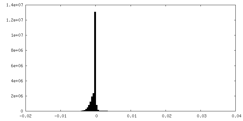
| Density Histograms |
 Z
Z Y
Y X
X


-Half map: #2
| File | emd_10617_half_map_1.map | ||||||||||||
|---|---|---|---|---|---|---|---|---|---|---|---|---|---|
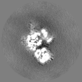
| Projections & Slices |
| ||||||||||||
| Density Histograms |


-Half map: #1
| File | emd_10617_half_map_2.map | ||||||||||||
|---|---|---|---|---|---|---|---|---|---|---|---|---|---|
| Projections & Slices |
| ||||||||||||
| Density Histograms |


- Sample components
-Entire : BBSome core complex
| Entire | Name: BBSome core complex |
|---|---|
| Components |
|
-Supramolecule #1: BBSome core complex
| Supramolecule | Name: BBSome core complex / type: complex / ID: 1 / Parent: 0 / Macromolecule list: all Details: BBSome core complex containing BBS1,4, 8, 9 and 18. BBS5 was also present in the sample preparation, but was only visible in a subset of particles (see related entry) |
|---|---|
| Source (natural) | Organism: Homo sapiens (human) |
-Macromolecule #1: Bardet-Biedl syndrome 1 protein
| Macromolecule | Name: Bardet-Biedl syndrome 1 protein / type: protein_or_peptide / ID: 1 / Number of copies: 1 / Enantiomer: LEVO |
|---|---|
| Source (natural) | Organism: Homo sapiens (human) |
| Molecular weight | Theoretical: 65.159266 KDa |
| Recombinant expression | Organism:  Trichoplusia ni (cabbage looper) Trichoplusia ni (cabbage looper) |
| Sequence | String: MAAASSSDSD ACGAESNEAN SKWLDAHYDP MANIHTFSAC LALADLHGDG EYKLVVGDLG PGGQQPRLKV LKGPLVMTES PLPALPAAA ATFLMEQHEP RTPALALASG PCVYVYKNLR PYFKFSLPQL PPNPLEQDLW NQAKEDRIDP LTLKEMLESI R ETAEEPLS ...String: MAAASSSDSD ACGAESNEAN SKWLDAHYDP MANIHTFSAC LALADLHGDG EYKLVVGDLG PGGQQPRLKV LKGPLVMTES PLPALPAAA ATFLMEQHEP RTPALALASG PCVYVYKNLR PYFKFSLPQL PPNPLEQDLW NQAKEDRIDP LTLKEMLESI R ETAEEPLS IQSLRFLQLE LSEMEAFVNQ HKSNSIKRQT VITTMTTLKK NLADEDAVSC LVLGTENKEL LVLDPEAFTI LA KMSLPSV PVFLEVSGQF DVEFRLAAAC RNGNIYILRR DSKHPKYCIE LSAQPVGLIR VHKVLVVGST QDSLHGFTHK GKK LWTVQM PAAILTMNLL EQHSRGLQAV MAGLANGEVR IYRDKALLNV IHTPDAVTSL CFGRYGREDN TLIMTTRGGG LIIK ILKRT AVFVEGGSEV GPPPAQAMKL NVPRKTRLYV DQTLREREAG TAMHRAFQTD LYLLRLRAAR AYLQALESSL SPLST TARE PLKLHAVVQG LGPTFKLTLH LQNTSTTRPV LGLLVCFLYN EALYSLPRAF FKVPLLVPGL NYPLETFVES LSNKGI SDI IKVLVLREGQ SAPLLSAHVN MPGSEGLAAA UniProtKB: Bardet-Biedl syndrome 1 protein |
-Macromolecule #2: Bardet-Biedl syndrome 4 protein
| Macromolecule | Name: Bardet-Biedl syndrome 4 protein / type: protein_or_peptide / ID: 2 / Number of copies: 1 / Enantiomer: LEVO |
|---|---|
| Source (natural) | Organism: Homo sapiens (human) |
| Molecular weight | Theoretical: 59.46302 KDa |
| Recombinant expression | Organism: Trichoplusia ni (cabbage looper) |
| Sequence | String: MHHHHHHGPM AEERVATRTQ FPVSTESQKP RQKKAPEFPI LEKQNWLIHL HYIRKDYEAC KAVIKEQLQE TQGLCEYAIY VQALIFRLE GNIQESLELF QTCAVLSPQS ADNLKQVARS LFLLGKHKAA IEVYNEAAKL NQKDWEISHN LGVCYIYLKQ F NKAQDQLH ...String: MHHHHHHGPM AEERVATRTQ FPVSTESQKP RQKKAPEFPI LEKQNWLIHL HYIRKDYEAC KAVIKEQLQE TQGLCEYAIY VQALIFRLE GNIQESLELF QTCAVLSPQS ADNLKQVARS LFLLGKHKAA IEVYNEAAKL NQKDWEISHN LGVCYIYLKQ F NKAQDQLH NALNLNRHDL TYIMLGKIHL LEGDLDKAIE VYKKAVEFSP ENTELLTTLG LLYLQLGIYQ KAFEHLGNAL TY DPTNYKA ILAAGSMMQT HGDFDVALTK YRVVACAVPE SPPLWNNIGM CFFGKKKYVA AISCLKRANY LAPFDWKILY NLG LVHLTM QQYASAFHFL SAAINFQPKM GELYMLLAVA LTNLEDIENA KRAYAEAVHL DKCNPLVNLN YAVLLYNQGE KKNA LAQYQ EMEKKVSLLK DNSSLEFDSE MVEMAQKLGA ALQVGEALVW TKPVKDPKSK HQTTSTSKPA SFQQPLGSNQ ALGQA MSSA AAYRTLPSGA GGTSQFTKPP SLPLEPEPAV ESSPTETSEQ IREK UniProtKB: Bardet-Biedl syndrome 4 protein |
-Macromolecule #3: Tetratricopeptide repeat domain 8 isoform 2
| Macromolecule | Name: Tetratricopeptide repeat domain 8 isoform 2 / type: protein_or_peptide / ID: 3 / Number of copies: 1 / Enantiomer: LEVO |
|---|---|
| Source (natural) | Organism: Homo sapiens (human) |
| Molecular weight | Theoretical: 58.702539 KDa |
| Recombinant expression | Organism: Trichoplusia ni (cabbage looper) |
| Sequence | String: MDYKDDDDKA GPMSSEMEPL LLAWSYFRRR KFQLCADLCT QMLEKSPYDQ AAWILKARAL TEMVYIDEID VDQEGIAEMM LDENAIAQV PRPGTSLKLP GTNQTGGPSQ AVRPITQAGR PITGFLRPST QSGRPGTMEQ AIRTPRTAYT ARPITSSSGR F VRLGTASM ...String: MDYKDDDDKA GPMSSEMEPL LLAWSYFRRR KFQLCADLCT QMLEKSPYDQ AAWILKARAL TEMVYIDEID VDQEGIAEMM LDENAIAQV PRPGTSLKLP GTNQTGGPSQ AVRPITQAGR PITGFLRPST QSGRPGTMEQ AIRTPRTAYT ARPITSSSGR F VRLGTASM LTSPDGPFIN LSRLNLTKYS QKPKLAKALF EYIFHHENDV KTALDLAALS TEHSQYKDWW WKVQIGKCYY RL GMYREAE KQFKSALKQQ EMVDTFLYLA KVYVSLDQPV TALNLFKQGL DKFPGEVTLL CGIARIYEEM NNMSSAAEYY KEV LKQDNT HVEAIACIGS NHFYSDQPEI ALRFYRRLLQ MGIYNGQLFN NLGLCCFYAQ QYDMTLTSFE RALSLAENEE EAAD VWYNL GHVAVGIGDT NLAHQCFRLA LVNNNNHAEA YNNLAVLEMR KGHVEQARAL LQTASSLAPH MYEPHFNFAT ISDKI GDLQ RSYVAAQKSE AAFPDHVDTQ HLIKQLRQHF AML UniProtKB: Tetratricopeptide repeat domain 8 isoform 2 |
-Macromolecule #4: Protein PTHB1
| Macromolecule | Name: Protein PTHB1 / type: protein_or_peptide / ID: 4 / Number of copies: 1 / Enantiomer: LEVO |
|---|---|
| Source (natural) | Organism: Homo sapiens (human) |
| Molecular weight | Theoretical: 99.383914 KDa |
| Recombinant expression | Organism: Trichoplusia ni (cabbage looper) |
| Sequence | String: MSLFKARDWW STILGDKEEF DQGCLCLANV DNSGNGQDKI IVGSFMGYLR IFSPHPAKTG DGAQAEDLLL EVDLRDPVLQ VEVGKFVSG TEMLHLAVLH SRKLCVYSVS GTLGNVEHGN QCQMKLMYEH NLQRTACNMT YGSFGGVKGR DLICIQSMDG M LMVFEQES ...String: MSLFKARDWW STILGDKEEF DQGCLCLANV DNSGNGQDKI IVGSFMGYLR IFSPHPAKTG DGAQAEDLLL EVDLRDPVLQ VEVGKFVSG TEMLHLAVLH SRKLCVYSVS GTLGNVEHGN QCQMKLMYEH NLQRTACNMT YGSFGGVKGR DLICIQSMDG M LMVFEQES YAFGRFLPGF LLPGPLAYSS RTDSFLTVSS CQQVESYKYQ VLAFATDADK RQETEQQKLG SGKRLVVDWT LN IGEQALD ICIVSFNQSA SSVFVLGERN FFCLKDNGQI RFMKKLDWSP SCFLPYCSVS EGTINTLIGN HNNMLHIYQD VTL KWATQL PHIPVAVRVG CLHDLKGVIV TLSDDGHLQC SYLGTDPSLF QAPNVQSREL NYDELDVEMK ELQKIIKDVN KSQG VWPMT EREDDLNVSV VVSPNFDSVS QATDVEVGTD LVPSVTVKVT LQNRVILQKA KLSVYVQPPL ELTCDQFTFE FMTPD LTRT VSFSVYLKRS YTPSELEGNA VVSYSRPTDR NPDGIPRVIQ CKFRLPLKLI CLPGQPSKTA SHKITIDTNK SPVSLL SLF PGFASQSDDD QVNVMGFHFL GGARITVLAS KTSQRYRIQS EQFEDLWLIT NELILRLQEY FEKQGVKDFA CSFSGSI PL QEYFELIDHH FELRINGEKL EELLSERAVQ FRAIQRRLLA RFKDKTPAPL QHLDTLLDGT YKQVIALADA VEENQGNL F QSFTRLKSAT HLVILLIALW QKLSADQVAI LEAAFLPLQE DTQELGWEET VDAAISHLLK TCLSKSSKEQ ALNLNSQLN IPKDTSQLKK HITLLCDRLS KGGRLCLSTD AAAPQTMVMP GGCTTIPESD LEERSVEQDS TELFTNHRHL TAETPRPEVS PLQGVSE UniProtKB: Protein PTHB1 |
-Macromolecule #5: BBSome-interacting protein 1
| Macromolecule | Name: BBSome-interacting protein 1 / type: protein_or_peptide / ID: 5 / Number of copies: 1 / Enantiomer: LEVO |
|---|---|
| Source (natural) | Organism: Homo sapiens (human) |
| Molecular weight | Theoretical: 15.430824 KDa |
| Recombinant expression | Organism: Trichoplusia ni (cabbage looper) |
| Sequence | String: MASWSHPQFE KGSAGSAAGS GAGWSHPQFE KGAGLEVLFQ GPKRAEFMLK AAAKRPELSG KNTISNNSDM AEVKSMFREV LPKQGPLFV EDIMTMVLCK PKLLPLKSLT LEKLEKMHQA AQNTIRQQEM AEKDQRQITH UniProtKB: BBSome-interacting protein 1 |
-Experimental details
-Structure determination
| Method | cryo EM |
|---|---|
 Processing Processing | single particle reconstruction |
| Aggregation state | particle |
-Sample preparation
| Concentration | 0.08 mg/mL | ||||||||
|---|---|---|---|---|---|---|---|---|---|
| Buffer | pH: 7.5 Component:
| ||||||||
| Grid | Model: UltrAuFoil / Material: GOLD / Mesh: 300 / Pretreatment - Type: GLOW DISCHARGE / Pretreatment - Time: 60 sec. / Pretreatment - Atmosphere: AIR | ||||||||
| Vitrification | Cryogen name: ETHANE / Chamber humidity: 100 % / Chamber temperature: 286 K / Instrument: FEI VITROBOT MARK III Details: double blot with 2 minutes incubation after first sample application. | ||||||||
| Details | The sample was cross linked with 0.5% glutaraldehyde |
- Electron microscopy
| Microscope | FEI TITAN KRIOS |
|---|---|
| Specialist optics | Phase plate: VOLTA PHASE PLATE / Energy filter - Slit width: 20 eV |
| Image recording | Film or detector model: GATAN K2 SUMMIT (4k x 4k) / Detector mode: COUNTING / Digitization - Frames/image: 1-50 / Number real images: 15266 / Average exposure time: 15.0 sec. / Average electron dose: 67.0 e/Å2 |
| Electron beam | Acceleration voltage: 300 kV / Electron source:  FIELD EMISSION GUN FIELD EMISSION GUN |
| Electron optics | Calibrated defocus max: 1.0 µm / Calibrated defocus min: 0.3 µm / Illumination mode: OTHER / Imaging mode: BRIGHT FIELD / Cs: 2.7 mm |
| Sample stage | Specimen holder model: FEI TITAN KRIOS AUTOGRID HOLDER / Cooling holder cryogen: NITROGEN |
| Experimental equipment |  Model: Titan Krios / Image courtesy: FEI Company |
