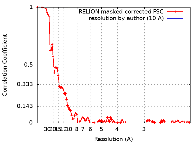
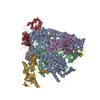
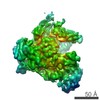
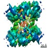
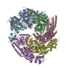
Journal: Nat Commun / Year: 2020 Title: Structure and mechanism of the Nap adhesion complex from the human pathogen Mycoplasma genitalium. Authors: David Aparicio / Margot P Scheffer / Marina Marcos-Silva / David Vizarraga / Lasse Sprankel / Mercè Ratera / Miriam S Weber / Anja Seybert / Sergi Torres-Puig / Luis Gonzalez-Gonzalez / ...Authors: David Aparicio / Margot P Scheffer / Marina Marcos-Silva / David Vizarraga / Lasse Sprankel / Mercè Ratera / Miriam S Weber / Anja Seybert / Sergi Torres-Puig / Luis Gonzalez-Gonzalez / Julian Reitz / Enrique Querol / Jaume Piñol / Oscar Q Pich / Ignacio Fita / Achilleas S Frangakis / Abstract: Mycoplasma genitalium is a human pathogen adhering to host target epithelial cells and causing urethritis, cervicitis and pelvic inflammatory disease. Essential for infectivity is a transmembrane ...Mycoplasma genitalium is a human pathogen adhering to host target epithelial cells and causing urethritis, cervicitis and pelvic inflammatory disease. Essential for infectivity is a transmembrane adhesion complex called Nap comprising proteins P110 and P140. Here we report the crystal structure of P140 both alone and in complex with the N-terminal domain of P110. By cryo-electron microscopy (cryo-EM) and tomography (cryo-ET) we find closed and open Nap conformations, determined at 9.8 and 15 Å, respectively. Both crystal structures and the cryo-EM structure are found in a closed conformation, where the sialic acid binding site in P110 is occluded. By contrast, the cryo-ET structure shows an open conformation, where the binding site is accessible. Structural information, in combination with functional studies, suggests a mechanism for attachment and release of M. genitalium to and from the host cell receptor, in which Nap conformations alternate to sustain motility and guarantee infectivity.
History
Deposition
Aug 26, 2019
-
Header (metadata) release
Jun 24, 2020
-
Map release
Jun 24, 2020
-
Update
Jun 24, 2020
-
Current status
Jun 24, 2020
Processing site: PDBe / Status: Released
-
Structure visualization
Movie
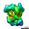
Surface view with section colored by density value
In the structure databanks used in Yorodumi, some data are registered as the other names, "COVID-19 virus" and "2019-nCoV". Here are the details of the virus and the list of structure data.
Jan 31, 2019. EMDB accession codes are about to change! (news from PDBe EMDB page)
EMDB accession codes are about to change! (news from PDBe EMDB page)
The allocation of 4 digits for EMDB accession codes will soon come to an end. Whilst these codes will remain in use, new EMDB accession codes will include an additional digit and will expand incrementally as the available range of codes is exhausted. The current 4-digit format prefixed with “EMD-” (i.e. EMD-XXXX) will advance to a 5-digit format (i.e. EMD-XXXXX), and so on. It is currently estimated that the 4-digit codes will be depleted around Spring 2019, at which point the 5-digit format will come into force.
The EM Navigator/Yorodumi systems omit the EMD- prefix.
Related info.:Q: What is EMD? / ID/Accession-code notation in Yorodumi/EM Navigator
Yorodumi is a browser for structure data from EMDB, PDB, SASBDB, etc.
This page is also the successor to EM Navigator detail page, and also detail information page/front-end page for Omokage search.
The word "yorodu" (or yorozu) is an old Japanese word meaning "ten thousand". "mi" (miru) is to see.
Related info.:EMDB / PDB / SASBDB / Comparison of 3 databanks / Yorodumi Search / Aug 31, 2016. New EM Navigator & Yorodumi / Yorodumi Papers / Jmol/JSmol / Function and homology information / Changes in new EM Navigator and Yorodumi
 Movie
Movie Controller

 Yorodumi
 Open data
 Basic information
 Map data
 Sample
 Mycoplasma genitalium G37 (bacteria)
 Authors
 Germany, 2 items
  Citation
 Structure visualization
 Movie viewer
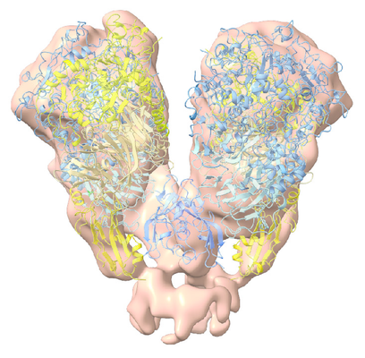
 Downloads & links
 emd_10260.png
 http://ftp.pdbj.org/pub/emdb/structures/EMD-10260


 Z (Sec.)
 Y (Row.)
 X (Col.)


 Sample components
 Processing
 Electron microscopy
 FIELD EMISSION GUN